Nidaan Dental
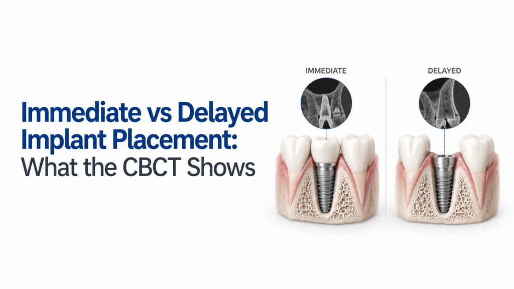
CBCT can help you see the three-dimensional conditions that influence whether immediate placement is realistic or whether allowing the extraction site to heal first may make the case more manageable. For an immediate implant, the scan can help assess the remaining facial bone, available bone for achieving primary stability, ridge dimensions, the relationship with adjacent roots and anatomical structures, and findings that may complicate treatment. For a delayed implant, CBCT can show the healed ridge and help determine the available bone and whether additional augmentation or another surgical approach may be required.
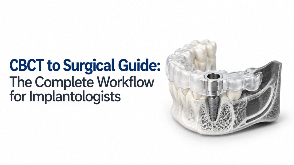
The CBCT to surgical guide workflow is a digital implant-planning process that combines three-dimensional CBCT information with intraoral or surface scan data to plan implant placement and produce a patient-specific surgical guide.
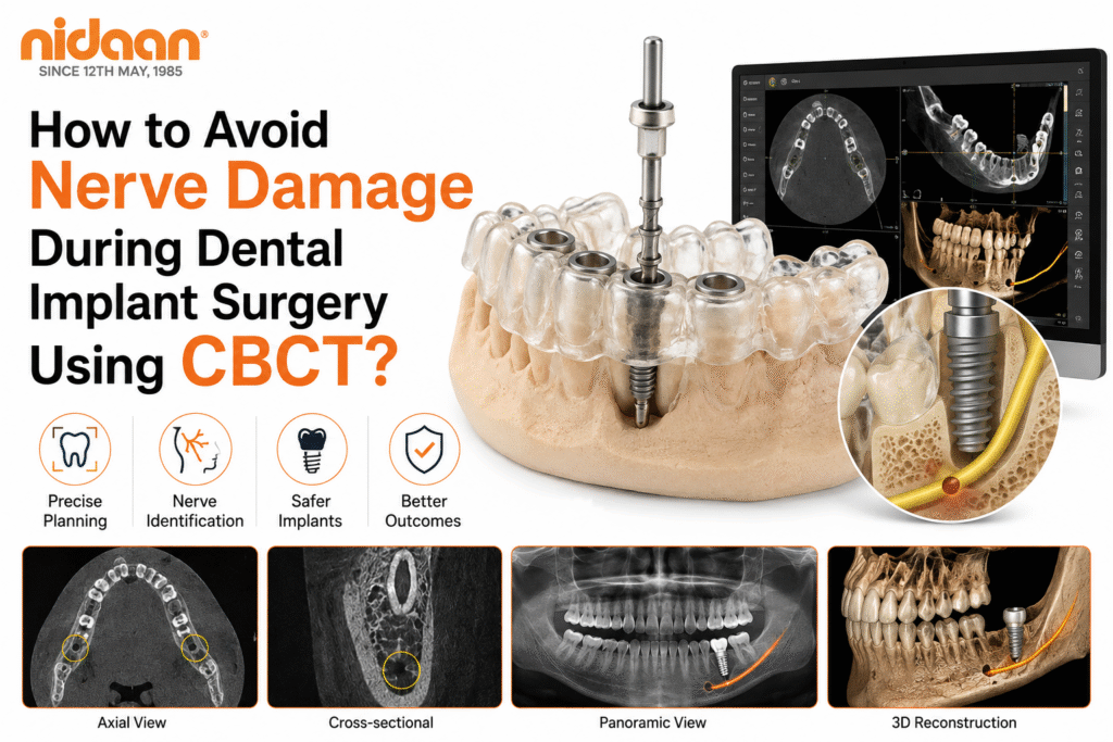
CBCT helps reduce the risk of inferior alveolar nerve injury during dental implant surgery by providing a three-dimensional view of the mandibular canal, mental foramen, anterior loop, and surrounding bone. This detailed visualization enables clinicians to plan implant position, angulation, and depth more accurately than conventional two-dimensional imaging. However, CBCT alone cannot prevent nerve damage. Safe implant placement depends on appropriate case selection, accurate interpretation of the scan, conservative safety margins, careful surgical execution, and continuous clinical judgment throughout treatment. A practical way to understand it is this: CBCT improves visibility. Digital planning improves precision. Clinical expertise protects the patient. When these elements work together, implant surgery becomes more predictable and significantly safer.
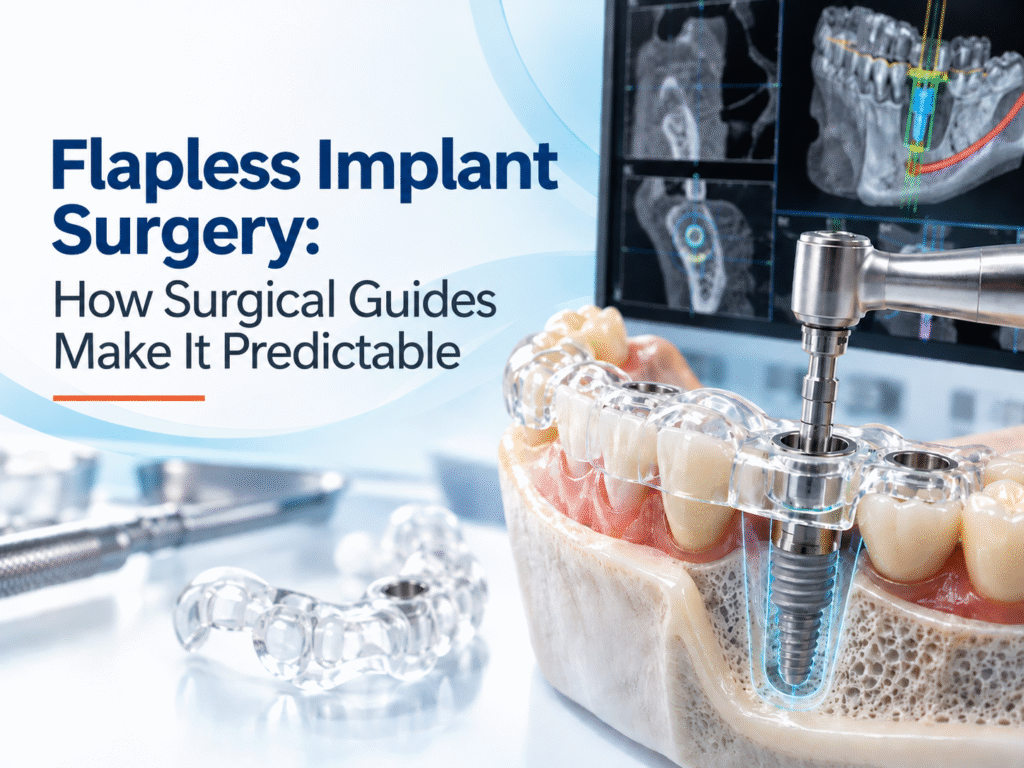
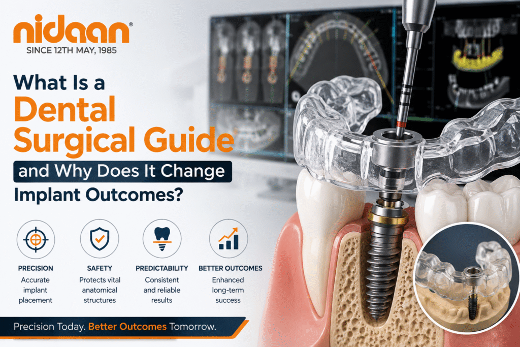
A dental surgical guide is a customized template, created from CBCT imaging and digital implant planning, that helps dentists place dental implants in the planned position before surgery. Instead of estimating implant location during the procedure, the guide directs the drills and implant according to the digital treatment plan, improving placement accuracy and supporting better prosthetic outcomes. However, a surgical guide is a precision aid, not a replacement for clinical expertise or comprehensive treatment planning.