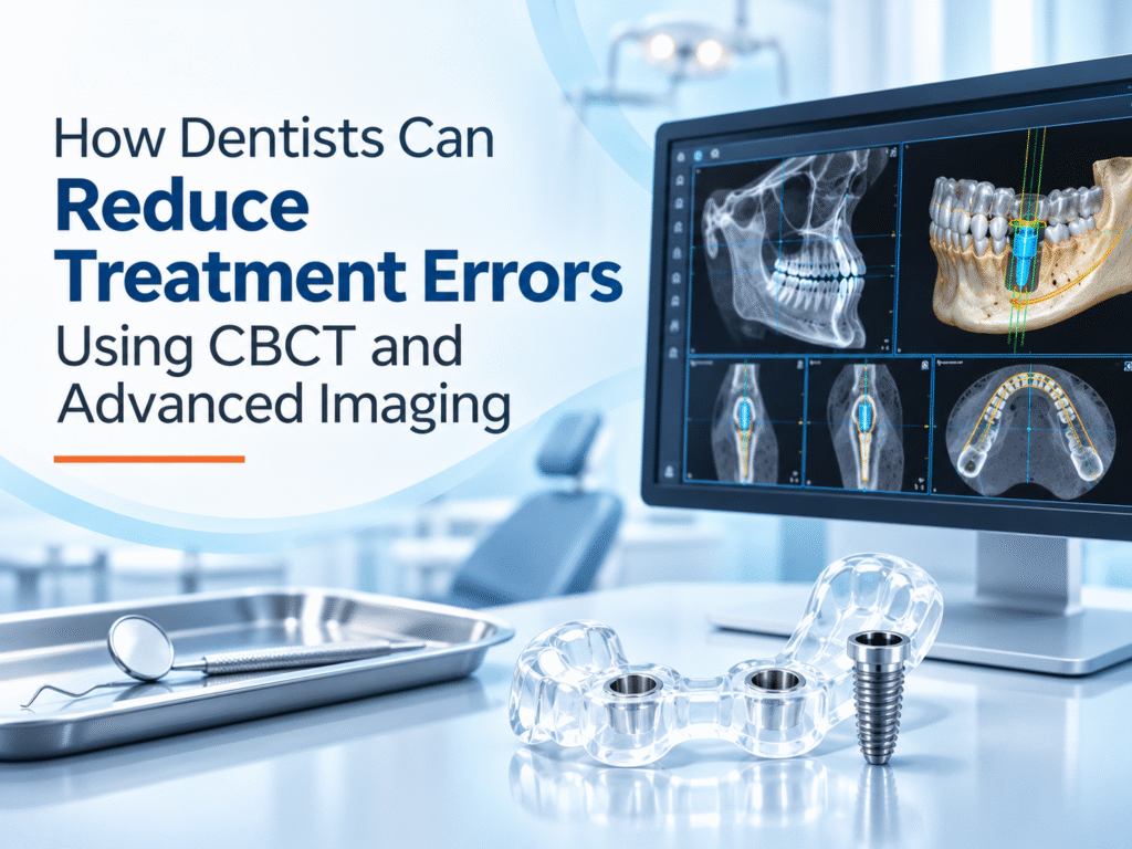

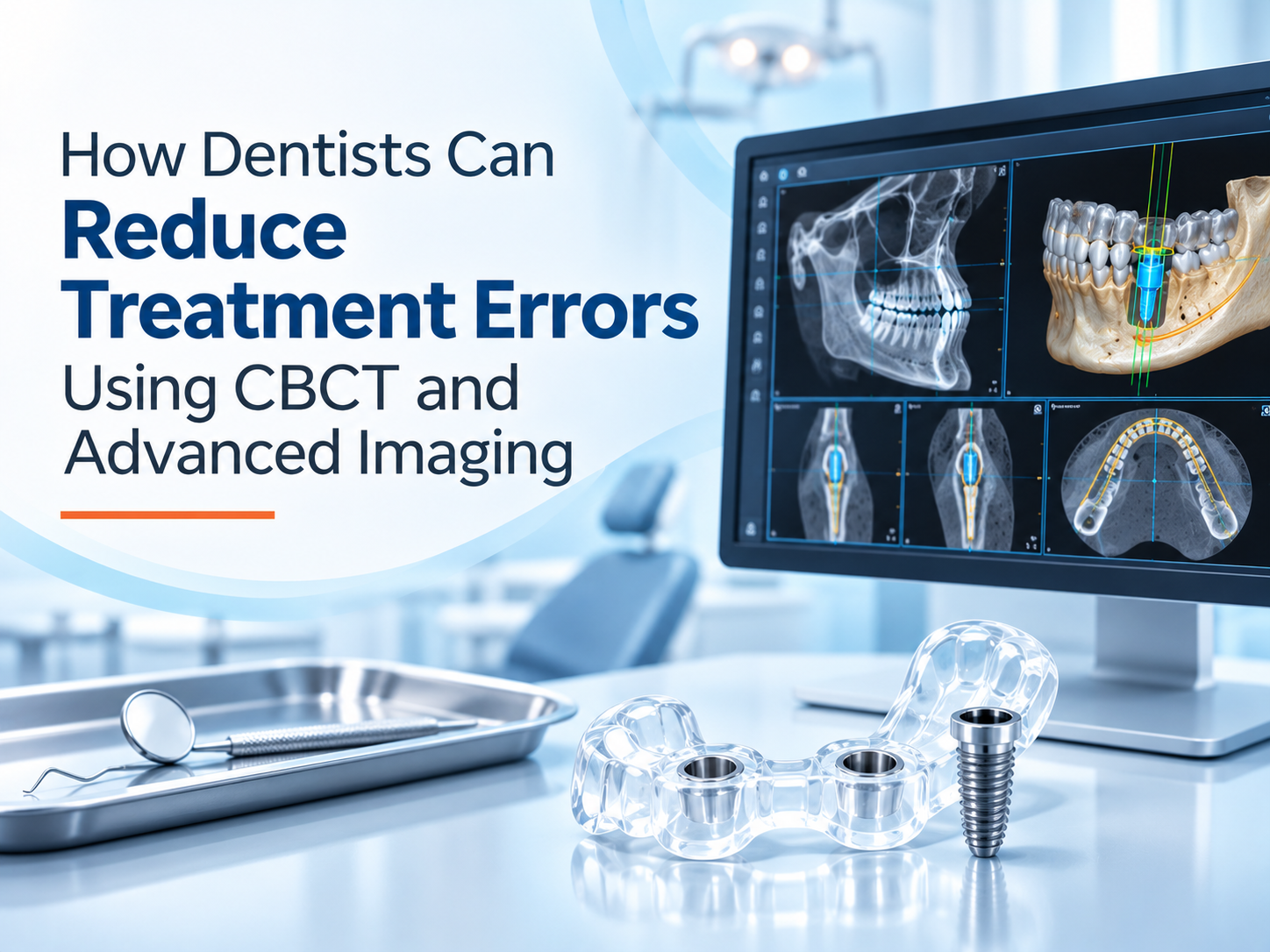
How Dentists Can Reduce Treatment Errors Using CBCT and Advanced Imaging

Dr. Akshay Shah
October 22, 2026
TL;DR
- Dental treatment errors can arise when important anatomical details, underlying pathology, or diagnostic findings are missed before treatment.
- OPG, intraoral radiographs, and CBCT help dentists evaluate different aspects of dental anatomy. Choosing the right modality matters more than ordering the most advanced scan.
- CBCT provides three-dimensional information that can support implant planning, complex endodontic assessment, impacted tooth evaluation, and selected surgical procedures.
- Structured radiology reporting helps dentists identify findings that may not be obvious during an initial image review.
- Digital implant planning and surgical guides can help transfer a planned implant position into surgery, but they cannot eliminate procedural risk.
- AI-assisted OPG screening can flag possible abnormalities for further assessment. It does not replace a formal radiological diagnosis or the dentist’s clinical judgment.
- A reliable error-reduction process combines clinical examination, justified imaging, image interpretation, treatment planning, and verification before treatment.
What if the biggest risk in a dental procedure is something the dentist cannot see?
Could a root canal fail because an additional canal was overlooked? Could an implant be planned too close to the mandibular nerve? Could an apparently straightforward extraction involve anatomy that changes the surgical approach?
These situations illustrate why diagnostic imaging matters before treatment begins. The objective is not simply to obtain clearer pictures. It is to identify findings that could change the clinical decision.
How Can Dentists Reduce Dental Treatment Errors Using Imaging?
Dentists can reduce the risk of treatment errors by selecting imaging based on the clinical question, reviewing relevant anatomy and pathology, obtaining specialist interpretation when needed, and using the findings to verify the treatment plan before proceeding.
Imaging is one part of clinical risk management. Patient history, examination findings, clinician experience, procedural technique, and follow-up remain essential.
The American Dental Association and American Academy of Oral and Maxillofacial Radiology’s 2026 consensus recommendations emphasize that radiographic examinations should follow clinical evaluation and be selected according to patient-specific needs.
Where Do Dental Treatment Errors Begin?
Some errors occur during a procedure. Others begin earlier, when a treatment plan is based on incomplete information.
A panoramic image may reveal the general position of an impacted tooth without fully establishing its three-dimensional relationship to the mandibular canal. A conventional radiograph may show a periapical lesion while leaving its full extent uncertain.
These limitations do not make two-dimensional imaging unreliable. They mean that every imaging method has a specific diagnostic role.
| Potential clinical risk | Imaging-related concern | How appropriate imaging helps |
| Implant positioned near a nerve | Incomplete assessment of canal location | CBCT helps evaluate the three-dimensional relationship |
| Missed root canal anatomy | Overlapping structures on 2D images | Selected small-field CBCT can clarify complex anatomy |
| Unexpected surgical difficulty | Unclear root position or adjacent structures | Additional views may improve surgical planning |
| Incorrect implant angulation | Incomplete assessment of available bone | CBCT supports prosthetically driven digital planning |
| Missed pathology | Findings overlooked during image review | Systematic interpretation and specialist reporting support detection |
The goal is to recognize uncertainty before it becomes a treatment complication.
How CBCT Helps Dentists Make Safer Treatment Decisions
1. Identifying Critical Anatomy Before Implant Surgery
Implant planning requires more than measuring the apparent height of available bone.
Dentists must consider ridge width, bone contours, the mandibular canal, mental foramen, maxillary sinus, and the planned prosthetic position.
CBCT provides cross-sectional views that can reveal anatomical relationships unavailable from a single panoramic projection.
For example, a mandibular implant site may appear to have sufficient vertical bone on an OPG. CBCT may reveal a lingual undercut or a narrower ridge than expected. That finding could change the implant dimensions, position, or surgical approach.
Clinical value: Better anatomical assessment helps dentists identify risks and adjust the plan before drilling.
2. Investigating Complex Root Canal Anatomy
Endodontic treatment depends on locating and treating the relevant root canal system.
Overlapping roots, unusual canal configurations, resorption, or suspected fractures may be difficult to evaluate using conventional radiographs alone.
In selected cases, limited-field CBCT can provide useful additional information.
It may help clinicians investigate:
- Suspected missed canals or complex root morphology.
- Persistent periapical disease after previous treatment.
- Internal or external root resorption.
- Possible root fractures when clinical findings and conventional images remain inconclusive.
CBCT is not necessary for every root canal. Its value is greatest when a specific unresolved question could change management.
3. Assessing Impacted Teeth and Surgical Relationships
An impacted mandibular third molar may lie close to the inferior alveolar canal. A two-dimensional image can suggest proximity, but it may not fully show the relationship between the roots and canal.
When clinically justified, CBCT can help the surgeon assess that relationship before selecting the treatment approach.
The same principle applies to selected impacted canines, complex extractions, and other procedures involving important anatomical structures.
CBCT improves the information available for planning. It does not remove the possibility of nerve injury or other surgical complications.
Choosing the Right Dental Imaging Modality
The most advanced scan is not automatically the best scan.
| Imaging modality | Main clinical role | Important limitation |
| Intraoral radiographs | Detailed assessment of individual teeth and surrounding structures | Two-dimensional projection |
| Bitewing radiographs | Evaluation of interproximal caries and crestal bone levels | Limited anatomical coverage |
| OPG | Broad overview of teeth, jaws, and related structures | Magnification, distortion, and overlapping anatomy |
| CBCT | Three-dimensional assessment of selected anatomical and pathological questions | Higher exposure than many conventional dental radiographs, with limited soft-tissue contrast |
The 2026 ADA and AAOMR recommendations advise using CBCT judiciously and considering existing images and lower-exposure alternatives.
Why Image Interpretation Matters as Much as Image Quality
A technically clear scan can still be clinically unhelpful if relevant findings are overlooked.
Dentists may focus on the immediate treatment site while other structures within the captured volume also require assessment.
A structured reporting process can support the review of relevant anatomy, pathology, and incidental findings.
The Role of Specialist CBCT Reporting
An Oral and Maxillofacial Radiologist can provide a dedicated interpretation of CBCT findings, particularly when a case involves complex anatomy, unusual pathology, or findings outside the referring dentist’s area of expertise.
This is where Nidaan Radtech fits into the workflow.
Nidaan Radtech provides CBCT teleradiology reporting by qualified Oral and Maxillofacial Radiologists, helping referring clinicians incorporate specialist imaging interpretation into treatment planning.
The radiology report supports clinical decisions. The referring dentist remains responsible for the final diagnosis and treatment plan.
From CBCT Imaging to Guided Implant Surgery
Identifying the correct implant position on a digital plan is one challenge. Reproducing that position during surgery is another.
A customized surgical guide can help transfer a digitally planned implant position into the operative setting.
How Nidaan UniGuide Supports This Process
Nidaan’s custom surgical guide workflow combines CBCT data with intraoral scan information.
The process involves:
- Capturing CBCT and intraoral scan data.
- Developing a patient-specific digital implant plan.
- Allowing the treating dentist to review and approve the proposed design.
- Fabricating a customized surgical guide.
- Using the guide during the planned implant procedure, subject to clinical suitability.
A surgical guide can support positioning and angulation, but its accuracy depends on the imaging, data alignment, design, fit, surgical technique, and other clinical factors.
It cannot compensate for an incorrect treatment plan.
Can AI Reduce Dental Diagnostic Errors?
AI can assist dentists by highlighting possible findings on dental radiographs, including suspected caries, bone changes, or periapical abnormalities.
Its practical role is to support screening and review, particularly when clinicians are assessing many images.
Nidaan offers an AI-assisted OPG screening report with its OPG imaging service.
The distinction matters: Nidaan’s AI OPG report is an automated preliminary screening output. It is not verified by a radiologist and does not constitute a formal radiological diagnosis.
Any flagged finding must be assessed by the treating dentist using the clinical examination and appropriate additional evidence.
AI should function as a supplementary review tool, not as an independent treatment decision-maker.
A Practical Imaging-Based Error-Reduction Workflow for Dental Clinics
Step 1: Define the clinical question.
Identify the uncertainty that imaging needs to resolve. Avoid ordering scans without a clear diagnostic purpose.
Step 2: Review existing clinical evidence.
Examine the patient, review medical and dental history, and assess previously obtained radiographs.
Step 3: Select the appropriate modality.
Use conventional imaging when sufficient. Consider CBCT when additional three-dimensional information is expected to affect diagnosis or treatment.
Step 4: Interpret the full relevant imaging information.
Evaluate anatomical relationships and possible pathology. Seek specialist radiology reporting where appropriate.
Step 5: Verify the treatment plan.
Confirm the planned procedure against imaging findings, clinical examination, and patient-specific risk factors.
Step 6: Communicate and document.
Explain relevant risks, alternatives, and limitations to the patient. Document the diagnostic reasoning and final plan.
Step 7: Reassess when findings change.
New symptoms, unexpected procedural findings, or changes in the clinical condition may require further evaluation.
This approach turns imaging into a clinical decision process rather than an isolated diagnostic appointment.
Advanced Dental Imaging Solutions for Dentists in Pune
For clinics seeking an imaging partner, the priority should be dependable diagnostic information that fits the treatment workflow.
Nidaan Dental supports dentists, clinics, and hospitals through complementary imaging and planning services.
| Nidaan service | How it supports clinical workflows |
| OPG and CBCT imaging | Provides 2D and 3D imaging for appropriate diagnostic and treatment-planning needs |
| Nidaan Radtech | Offers specialist CBCT interpretation and structured radiology reporting |
| Nidaan UniGuide | Supports digital implant planning and fabrication of customized surgical guides |
| AI-assisted OPG screening | Provides preliminary automated findings for clinician review |
With over four decades of experience and a network of more than 1,200 dentist partners, Nidaan focuses on supporting dental professionals with imaging, interpretation, and planning services.
Make Imaging Part of Your Treatment Safety Workflow
Whether you are assessing a complex implant site, investigating difficult anatomy, or planning a guided procedure, the right diagnostic information can help you make a more informed clinical decision.
Work with Nidaan Dental for OPG and CBCT imaging, specialist reporting, and customized implant surgical guides in Pune.
Explore Nidaan Dental Services | Contact Nidaan Dental
Conclusion
Reducing dental treatment errors starts before the procedure.
The most useful imaging workflow is one that answers a clear clinical question, identifies relevant risks, and gives the dentist enough information to select an appropriate treatment approach.
CBCT, specialist radiology reporting, digital planning, and surgical guides can strengthen that process when used for the right indications.
The technology supports the decision. Clinical judgment remains central to patient safety.
Frequently Asked Questions
1. How can dentists reduce treatment errors using advanced imaging?
Dentists can use clinically justified imaging to identify anatomical risks, investigate uncertain findings, support treatment planning, and verify critical details before a procedure. Imaging should complement examination and clinical judgment.
2. Does CBCT prevent dental treatment errors?
No. CBCT provides additional three-dimensional diagnostic information that can help clinicians recognize risks and plan treatment. It cannot guarantee that complications or errors will not occur.
3. Is CBCT better than OPG for implant planning?
CBCT provides three-dimensional information about bone dimensions and nearby anatomical structures. OPG offers a broad two-dimensional overview. The appropriate modality depends on the clinical question and the information required for treatment planning.
4. Can CBCT help prevent nerve injury during implant surgery?
CBCT can help dentists identify the mandibular canal and related anatomical structures before implant placement. This supports risk assessment and planning but does not eliminate the possibility of nerve injury.
5. Is CBCT necessary for every root canal?
No. Conventional radiographs are sufficient for many endodontic cases. CBCT may be useful when complex anatomy, persistent disease, suspected fractures, or other specific concerns remain unresolved.
6. How does CBCT help with guided implant surgery?
CBCT provides three-dimensional anatomical data that can be combined with digital dental scans to plan implant positioning and design a patient-specific surgical guide.
7. What is the benefit of a CBCT radiology report?
A specialist report provides structured interpretation of imaging findings and can help the referring dentist evaluate complex anatomy or pathology. It supports, rather than replaces, the dentist’s final clinical decisions.
8. Can AI detect dental problems accurately?
AI may identify possible abnormalities for further review, but its performance varies by system and clinical condition. Findings require professional assessment and should not be treated as an independent diagnosis.
9. Does advanced dental imaging increase radiation exposure?
Radiation exposure depends on the modality, equipment, scan settings, and area examined. CBCT generally involves more exposure than many conventional dental radiographs, so its use should be justified and optimized for the clinical need.
10. Where can dentists access OPG, CBCT, and surgical guide services in Pune?
Nidaan Dental provides OPG and CBCT imaging in Pune, along with Nidaan Radtech reporting and customized implant surgical guides through Nidaan UniGuide. Dentists can review the available services on the official Nidaan Dental website.