
How to Avoid Nerve Damage in Implant Surgery Using CBCT
September 23, 2026
TL;DR
- Inferior alveolar nerve injury is one of the important complications to consider during mandibular implant surgery.
- CBCT provides three-dimensional views of the mandibular canal, mental foramen, anterior loop and other anatomical structures that may be difficult to assess on 2D radiographs.
- The safest approach is not simply to “take a CBCT.” It is to use CBCT as part of a structured risk-management workflow.
- Nerve mapping should include the mandibular canal and relevant anatomical variations, not just a single measurement on one CBCT slice.
- Implant position should be planned from both the anatomical and prosthetic perspectives.
- There is no universal implant-to-nerve distance that guarantees safety for every patient.
- Surgical guides can improve transfer of the planned implant position, but they cannot correct an incorrect plan or eliminate surgical risk.
- CBCT should be prescribed when three-dimensional information is needed to answer a clinical question or influence treatment planning.
- If the clinical findings do not match the CBCT-based plan, the surgeon should stop, reassess and modify the approach rather than blindly follow the digital plan.
- CBCT reduces uncertainty. It does not eliminate risk.
Direct Answer: How Does CBCT Help Avoid Nerve Damage During Implant Surgery?
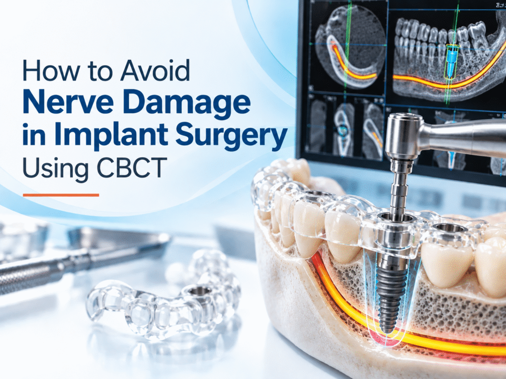
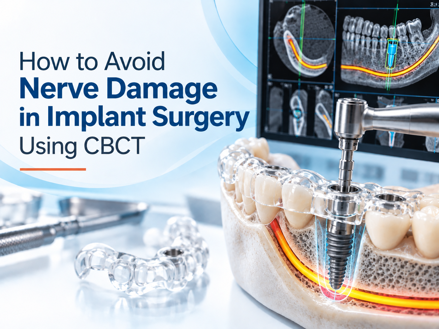
CBCT helps reduce the risk of nerve injury by showing the three-dimensional relationship between the planned implant site and important neural structures such as the inferior alveolar nerve, mental foramen and anterior loop.
Unlike a panoramic or periapical radiograph, CBCT allows clinicians to assess the anatomy in multiple planes. This can reveal the buccolingual position of the mandibular canal, anatomical variations, ridge morphology and the relationship between the planned implant and the nerve.
But CBCT does not prevent nerve damage by itself.
Safe implant surgery requires a combination of accurate imaging, careful nerve mapping, patient-specific planning, appropriate safety margins, surgical-guide verification and clinical judgment during the procedure.
Why Nerve Damage Matters in Implant Surgery
The inferior alveolar nerve travels through the mandibular canal and provides sensation to the lower teeth, lower lip and chin. An implant, osteotomy or other surgical intervention that affects the nerve can result in altered sensation.
Possible symptoms include:
- Numbness or reduced sensation
- Tingling or pins-and-needles sensations
- Burning or electric-shock-like pain
- Altered sensation in the lower lip or chin
- Hypersensitivity
- Changes in temperature or touch perception
- Persistent neuropathic pain in more severe cases
The clinical problem is not limited to placing an implant directly into the nerve canal. Nerve-related complications can also involve the osteotomy, surgical instruments, grafting procedures, postoperative swelling or other aspects of treatment.
That is why implant planning should identify the relevant anatomy before surgery rather than relying only on what becomes apparent during the procedure.
What Should CBCT Show Before a Mandibular Implant?
A useful CBCT assessment goes beyond measuring vertical bone height.
Inferior Alveolar Nerve and Mandibular Canal
The mandibular canal should be traced through multiple planes to understand its course relative to the planned implant.
The clinician needs to assess more than whether the canal appears “below” the proposed implant. Its buccolingual position, vertical relationship and surrounding cortical boundaries can affect the available surgical space.
CBCT is particularly useful when the implant site is close to the canal or when conventional imaging does not provide enough information for treatment planning.
Mental Foramen and Anterior Loop
The mental foramen is another important landmark, particularly when planning implants in the premolar region.
The anterior loop refers to the portion of the mental nerve that may extend anteriorly before exiting through the mental foramen. Its location and extent can vary between individuals.
If the anterior loop is not identified, an implant planned from a 2D image may be positioned closer to the nerve than expected.
CBCT allows the clinician to evaluate this anatomy in three dimensions and modify implant position when necessary.
Anatomical Variations
Not every mandibular canal follows the same path.
Planning may need to account for:
- Bifid or trifid mandibular canals
- Accessory mental foramina
- Retromolar canals
- Accessory neurovascular pathways
- Lingual or buccal anatomical concavities
- Variations in ridge width and morphology
These findings are one reason a single measurement should not be treated as the entire safety assessment.
Why a Panoramic X-Ray May Not Be Enough
A panoramic image provides useful information, but it compresses three-dimensional anatomy into a two-dimensional image.
This can make it difficult to determine:
- The buccolingual position of the mandibular canal
- The exact vertical relationship between the canal and the ridge
- Lingual or buccal concavities
- The course of an anterior loop
- Accessory mental foramina
- The relationship between the planned implant and the nerve in cross-section
CBCT adds multiplanar and three-dimensional information when that additional detail is clinically necessary.
That does not mean every implant requires CBCT. Imaging should be justified according to the diagnostic question, with the smallest appropriate field of view and exposure compatible with the clinical need.
The CBCT Safety Workflow for Implant Surgery
A safer workflow starts before implant placement.
1. Identify the Clinical Risk
First determine whether the implant site presents a meaningful anatomical question.
Posterior mandibular implants close to the inferior alveolar canal, premolar implants near the mental foramen or cases where conventional imaging is ambiguous may require three-dimensional assessment.
The purpose of the scan should be clear: What information does the clinician need that cannot be adequately obtained from conventional imaging?
2. Trace the Nerve in Multiple Planes
Do not assess the nerve from a single panoramic reconstruction or one CBCT slice.
The mandibular canal should be followed through the available planes so that its course, position and relationship with the proposed implant can be understood.
The same principle applies to the mental foramen and anterior loop.
3. Assess the Whole Implant Site
Nerve safety is only one part of implant planning.
The clinician should also evaluate:
- Bone height and width
- Ridge morphology
- Cortical boundaries
- Adjacent teeth and roots
- Implant diameter and length
- Prosthetic position
- Available space for the planned restoration
An implant that is technically distant from the nerve can still be poorly positioned if the restorative and anatomical objectives are ignored.
4. Use an Individualized Safety Margin
One of the most common mistakes is treating a specific number as a universal guarantee.
Many clinical discussions refer to planning buffers around the inferior alveolar nerve, but there is no single distance that guarantees nerve protection for every patient.
Measurement uncertainty, image quality, anatomical variation, implant dimensions and surgical technique all matter.
The safety margin should therefore be determined case by case rather than reduced to a fixed rule.
5. Verify the Digital Surgical Plan
When guided surgery is used, the guide should be treated as one component of the safety workflow.
The planned implant position needs to be reviewed before the guide is manufactured and used. The clinician should confirm that the proposed trajectory remains appropriate for the patient’s anatomy and restorative objectives.
A surgical guide improves the transfer of a plan. It does not make an incorrect plan safe.
6. Reassess During Surgery
The CBCT represents the patient’s anatomy as captured during imaging. Clinical findings during surgery still matter.
If the anatomy, guide fit or surgical situation does not correspond with the planned procedure, the clinician should reassess rather than continue simply because the digital plan has already been approved.
This is one of the most important distinctions between using technology to support treatment and allowing technology to dictate treatment.
A Practical Example: When CBCT Changes the Implant Plan
Consider a patient who needs an implant in the lower premolar region.
A panoramic radiograph appears to show sufficient vertical bone. The clinician then obtains CBCT imaging because the planned implant site is close to the mental foramen.
The three-dimensional assessment reveals that:
- The mental foramen is positioned more anteriorly than initially expected.
- The anterior loop extends into the originally planned implant area.
- The available clearance from the mandibular canal is less than estimated from the panoramic image.
Instead of proceeding with the original plan, the clinician can reconsider implant dimensions, angulation or position and establish a more conservative relationship with the nerve.
The important point is that CBCT did not prevent nerve injury automatically. It provided information that allowed the treatment plan to change before surgery.
Can Guided Implant Surgery Reduce Nerve Risk?
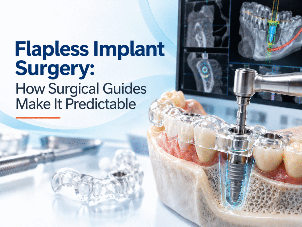
Guided surgery can help transfer a carefully planned implant position, angulation and drilling path to the surgical site.
In a CBCT-based guided workflow, the digital plan can be used to create a patient-specific surgical guide. This can improve consistency in positioning and provide an additional layer of control during implant placement.
But guided surgery does not guarantee nerve protection.
A guide can still be affected by:
- Incorrect digital planning
- Inaccurate nerve mapping
- CBCT or registration errors
- Poor guide fit
- Guide movement during surgery
- Drill deviation
- Incorrect clinical execution
The guide should therefore be viewed as part of the risk-management process, not as a substitute for clinical expertise.
Common Myths About CBCT and Nerve Safety
| Myth | Evidence-based view |
|---|---|
| CBCT completely prevents nerve injury | CBCT improves three-dimensional visualization but cannot eliminate surgical risk. |
| A fixed 2 mm distance is always safe | Safety margins should be individualized according to anatomy and measurement uncertainty. |
| Guided surgery guarantees nerve protection | A guide improves transfer of the plan but cannot correct poor planning. |
| Every implant requires CBCT | CBCT should be used when 3D information is clinically necessary. |
| Automatic nerve tracing replaces clinician review | Automated or digital tools still require appropriate clinical and radiological interpretation. |
These distinctions matter because the purpose of CBCT is to reduce uncertainty and improve decision-making, not to create a false sense of absolute safety.
How Nidaan Supports CBCT-Based Implant Planning
Nidaan provides CBCT imaging for dental treatment planning, with its workflow focused on giving dentists three-dimensional information for clinical decision-making.
For cases where guided implant placement is appropriate, Nidaan UniGuide combines CBCT data with digital implant planning and customized surgical-guide fabrication. The workflow is designed to help transfer a carefully reviewed implant position to the surgical field.
The important distinction is that the technology supports the clinician’s plan. It does not replace the dentist’s responsibility for diagnosis, case selection, surgical execution or responding to unexpected findings.
Conclusion
Avoiding nerve damage during implant surgery starts before the first osteotomy.
CBCT can give the implantologist a much clearer three-dimensional view of the inferior alveolar nerve, mental foramen, anterior loop and surrounding anatomy. That information can reveal anatomical relationships that are difficult to appreciate on conventional two-dimensional imaging and can change the planned implant position before surgery.
But the safest approach is not simply “get a CBCT.”
It is to use CBCT as part of a complete workflow: identify the relevant anatomy, trace the nerve in multiple planes, recognize anatomical variations, plan the implant from both anatomical and prosthetic perspectives, establish an individualized safety margin, verify the surgical guide when used and remain prepared to modify the procedure when clinical findings require it.
CBCT reduces uncertainty. It does not eliminate risk. The value of three-dimensional imaging comes from how accurately that information is interpreted and how effectively it is incorporated into clinical decision-making.
FAQs
1. Can a CBCT scan completely prevent nerve damage during implant surgery?
No. CBCT improves visualization of the inferior alveolar nerve, mental foramen, anterior loop and surrounding anatomy, but it cannot eliminate the possibility of nerve injury. Safe implant surgery also depends on accurate interpretation, individualized planning, appropriate safety margins and careful surgical execution.
2. Why is CBCT useful for implants near the inferior alveolar nerve?
A conventional panoramic image provides a two-dimensional representation of the jaw. CBCT provides multiplanar and three-dimensional views, allowing the clinician to evaluate the mandibular canal’s position, surrounding bone, anatomical variations and the relationship between the planned implant and the nerve.
3. Does every dental implant require a CBCT scan?
No. CBCT should be prescribed when three-dimensional information is necessary to answer a specific clinical question or influence treatment planning. Straightforward cases may not always require CBCT if conventional imaging provides sufficient information.
4. What anatomical structures should be checked on CBCT before a mandibular implant?
Depending on the implant site, clinicians may assess the inferior alveolar canal, mental foramen, anterior loop, incisive canal, accessory mental foramina, bifid or trifid canals, ridge morphology, bone dimensions and lingual or buccal concavities.
5. What is the anterior loop of the mental nerve?
The anterior loop is an anatomical extension of the mental nerve that may extend forward before the nerve exits through the mental foramen. Its position varies between individuals. Identifying it on CBCT can help prevent an implant planned in the premolar region from being positioned too close to the nerve.
6. Is there a universally safe distance between an implant and the inferior alveolar nerve?
No. Although specific planning buffers are discussed in implant dentistry, there is no single distance that guarantees nerve protection in every patient. The appropriate margin depends on anatomy, image quality, implant dimensions, measurement uncertainty and surgical factors.
7. Can guided implant surgery prevent inferior alveolar nerve injury?
Guided surgery can improve the consistency with which a carefully planned implant position is transferred to the surgical site. It cannot compensate for inaccurate nerve mapping, poor planning, guide movement, registration errors or surgical deviation. It should be considered one part of a comprehensive safety workflow.
8. What happens if the CBCT shows that the implant is too close to the nerve?
The clinician may need to reconsider the implant’s position, angulation, dimensions or treatment approach. The appropriate response depends on the patient’s anatomy and treatment objectives. The key advantage of CBCT is that it can reveal the problem during planning, before the surgical procedure begins.
9. Can nerve damage happen even when the implant itself does not enter the mandibular canal?
Yes. Nerve-related complications are not limited to direct implant contact with the nerve. Osteotomy, surgical instruments, grafting, postoperative swelling and other surgical factors can also affect neural structures.
10. How does Nidaan UniGuide support safer implant planning?
Nidaan UniGuide combines CBCT-based three-dimensional information, digital implant planning and customized surgical-guide fabrication. It can help the clinician transfer a reviewed implant position to the surgical field. It remains a planning and surgical-support tool, not a replacement for clinical judgment.