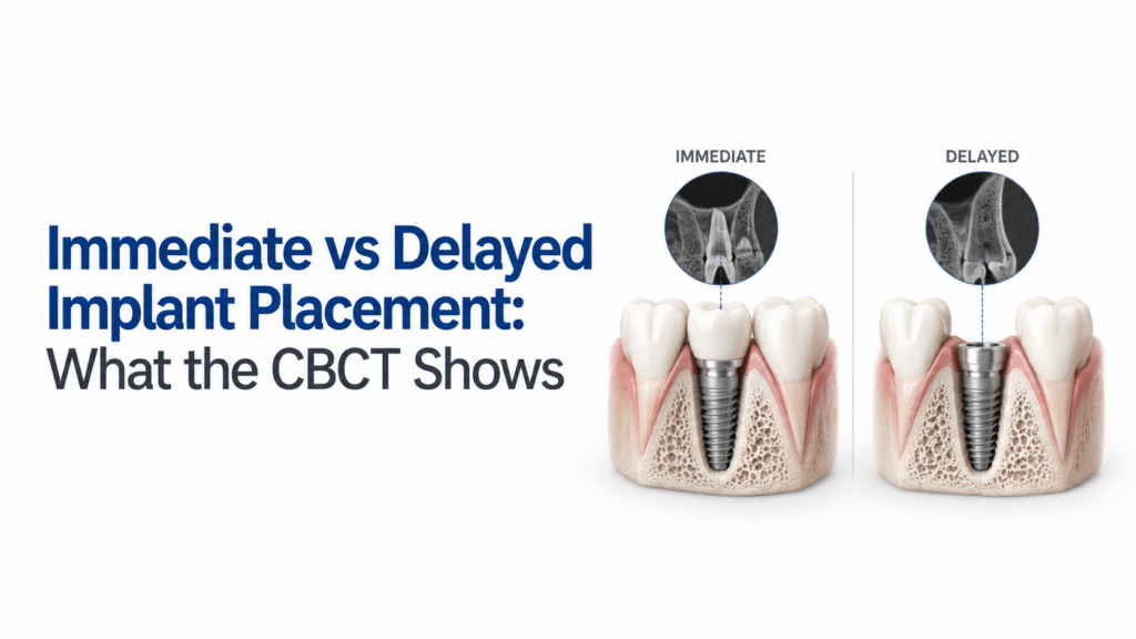

Immediate vs Delayed Implant Placement: What the CBCT Shows

Dr. Akshay Shah
August 26, 2026
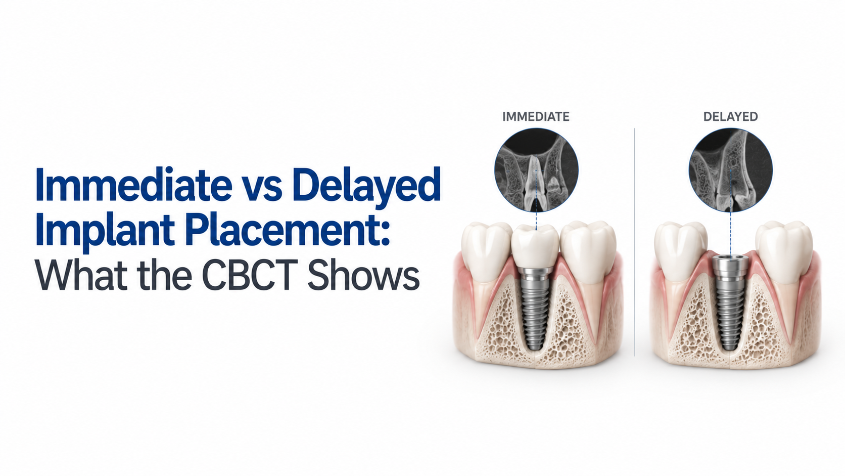
TL;DR
- Immediate implant placement means placing the implant at the time of extraction, while delayed placement is performed after further healing of the extraction site. These are different timing protocols and should not be confused with immediate loading.
- The key question is not whether immediate or delayed placement is universally better. It is what the patient’s anatomy and healing situation allow.
- CBCT helps the implantologist assess the three-dimensional anatomy that may be difficult to appreciate from 2D imaging alone.
- Before considering immediate placement, CBCT assessment should pay particular attention to the facial/buccal bone plate, available bone for primary stability, ridge dimensions, adjacent structures and possible pathology or defects.
- A thin or missing buccal plate can change the risk profile of an immediate case, particularly in esthetically demanding sites.
- CBCT can support treatment timing decisions, but it cannot replace the clinical examination, extraction assessment, soft-tissue evaluation or prosthetic plan.
- Evidence does not show a universal survival advantage for either approach. A 2023 meta-analysis reported 97.4% survival for immediate implants and 97.5% for delayed implants, with no statistically significant difference.
- A 2026 CBCT-based study of 60 grafted extraction sites found greater buccal bone preservation with delayed placement, while implant stability and 12-month survival were comparable between groups.
- The most useful CBCT question is therefore not “Which protocol wins?”, but “What does this anatomy tell me about the risks and requirements of each option?”
- CBCT makes the anatomical trade-offs more visible. The final timing decision still belongs to the treating clinician.
What Does CBCT Show in Immediate vs Delayed Implant Placement?
CBCT can help you see the three-dimensional conditions that influence whether immediate placement is realistic or whether allowing the extraction site to heal first may make the case more manageable.
For an immediate implant, the scan can help assess the remaining facial bone, available bone for achieving primary stability, ridge dimensions, the relationship with adjacent roots and anatomical structures, and findings that may complicate treatment. For a delayed implant, CBCT can show the healed ridge and help determine the available bone and whether additional augmentation or another surgical approach may be required.
So, CBCT does not choose immediate or delayed placement by itself. It gives you anatomical information that can make the timing decision more informed.
The International Team for Implantology (ITI) consensus recommends good-quality periapical imaging and CBCT for appropriate assessment of immediate placement sites, including evaluation of the facial bone plate and bone available for an ideal 3D implant position.
Immediate vs Delayed Implant Placement: What Is the Difference?
Immediate placement occurs on the same day as tooth extraction. Delayed placement occurs after the socket has undergone further healing. The ITI classification also distinguishes early placement from late placement, so “delayed” should not automatically be treated as one fixed waiting period.
The biological trade-off is straightforward. Immediate placement can shorten the time between extraction and implant placement, but the implant is being positioned in an extraction site whose anatomy is changing. Delaying treatment allows healing to occur first, which may make the ridge easier to assess and manage, although healing can also involve dimensional changes in the ridge.
Stephen T. Chen, MDSc, PhD, FRACDS, Clinical Associate Professor at the University of Melbourne and Past President of the ITI, describes the central challenge clearly: “The challenge lies in managing resorption of the alveolar ridge once a tooth is extracted.”
That is why timing is a clinical decision rather than a simple preference for faster or slower treatment.
What Should You Look for on CBCT Before Immediate Implant Placement?
1. Facial or Buccal Bone
The facial bone plate deserves close attention, particularly in the anterior maxilla. CBCT can reveal whether the facial plate is intact, thin or absent and how the planned implant position relates to it.
This matters because an implant cannot simply be placed according to the extraction socket’s existing shape. The implant position still needs to satisfy restorative and biological requirements.
One CBCT study examining 1,104 anterior maxillary teeth found mean buccal bone thickness of 0.55 ± 0.38 mm at one measurement point and 0.60 ± 0.30 mm at another. Missing buccal bone was also common in the studied population.
These numbers are useful for understanding why the facial plate deserves attention, but they should not be turned into a universal cutoff for every immediate case.
2. Bone Available for Primary Stability
Immediate placement requires sufficient bone in an appropriate three-dimensional position to achieve primary stability.
CBCT helps you look beyond the extraction socket itself. In some cases, the socket may appear suitable on a 2D image, while the 3D scan shows that the planned implant trajectory would leave inadequate supporting bone.
The question becomes: Where can the implant be positioned prosthetically and anatomically while still obtaining the stability required for the planned protocol?
The ITI consensus specifically lists sufficient bone for primary stability in an ideal 3D position among the radiographic criteria for immediate placement.
3. Ridge Dimensions and Defect Pattern
CBCT can show the width and height of the available ridge and reveal defects that may affect immediate placement.
Look at the buccal and palatal or lingual boundaries rather than judging the socket from one cross-section. A localized dehiscence, fenestration or loss of the facial plate can change the surgical plan.
This is particularly important because the evidence is not uniform across every defect pattern. A 10-year randomized clinical trial involving 40 patients with failing anterior teeth and buccal defects of at least 5 mm found no statistically significant differences in marginal bone levels, clinical outcomes, aesthetics or patient satisfaction between immediate placement with augmentation and delayed placement after ridge preservation.
The point is not that defects automatically rule out immediate placement. They change the planning conversation.
When CBCT Findings May Favor a Delayed Approach
There is no single CBCT measurement that automatically means “wait.” The decision depends on the entire case, including extraction findings, soft tissue, infection, restorative requirements and the ability to obtain predictable implant stability.
Still, delayed placement may become more attractive when the scan shows:
| CBCT finding | Why it matters |
| Significant facial/buccal bone deficiency | Immediate positioning may require additional management |
| Limited bone for primary stability | The planned implant position may not provide adequate support |
| Unfavorable ridge dimensions | Healing or augmentation may improve the available site |
| Complex anatomical relationship | Additional planning may be needed before placement |
| Pathology or concerning local findings | The underlying condition may influence timing |
| Poor prosthetically driven implant position | Immediate placement may compromise the restorative objective |
The 2026 CBCT-based study in the supplied research is a useful example of why timing cannot be reduced to survival alone. In 60 grafted extraction sites, delayed placement showed less buccal bone loss than immediate placement, with reported losses of 0.42 ± 0.28 mm versus 0.89 ± 0.34 mm. Yet implant stability, integration and 12-month survival were comparable, and both groups reported 100% survival at 12 months.
Does CBCT Show That Immediate or Delayed Implants Have Better Survival?
Not consistently.
The supplied research reports a 2023 systematic review and meta-analysis covering 10 studies, with 341 immediately placed implants and 359 delayed implants. Survival was 97.4% and 97.5%, respectively, and the pooled difference was not statistically significant.
A 2024 multicenter randomized clinical trial also found 95.8% survival in the immediate group and 92% in the delayed group at the reported follow-up, showing why individual studies can point in different directions.
So if you are using CBCT to answer “Which timing has the highest survival?”, you are asking the scan to answer a question it was not designed to answer. Its greater value is showing you why a particular site may or may not be suitable for one timing protocol.
A Practical CBCT Decision Framework
Before choosing immediate or delayed placement, review the case in this order:
- Assess the extraction site: What is likely to remain after atraumatic extraction?
- Review the facial plate: Is it intact, thin or deficient?
- Check three-dimensional ridge anatomy: Is there enough bone for the intended implant position?
- Assess primary stability potential: Can the implant be anchored in suitable native bone?
- Review adjacent anatomy: Are roots or other structures affecting the planned trajectory?
- Consider pathology and defects: Is there anything that changes the biological or surgical situation?
- Overlay the prosthetic plan: Does the ideal restorative position remain achievable?
- Compare timing options: Would immediate placement offer a clear clinical advantage, or would healing first make the case more predictable?
This approach keeps CBCT in its proper role. It informs the decision rather than deciding for you.
Immediate vs Delayed Implant Placement: The Bottom Line
There is no universal winner between immediate and delayed implant placement. The better question is whether the specific site supports the intended protocol.
CBCT is particularly useful when you need to understand the three-dimensional relationship between the extraction socket, facial and palatal or lingual bone, adjacent anatomy and the planned implant position. It can reveal limitations that are easy to underestimate on conventional radiographs.
The evidence also argues against blanket claims. Survival can be comparable between protocols, while bone changes and clinical requirements may differ by site and treatment approach.
For an implantologist, the practical takeaway is simple: use CBCT to understand the anatomy before deciding the timing, not to justify a timing decision you have already made.
Conclusion
Immediate and delayed implant placement are both established treatment approaches, but neither should be selected from a generic checklist of advantages and disadvantages. The anatomy of the individual extraction site changes the decision.
CBCT gives you a better view of that anatomy. It can show the facial bone plate, three-dimensional ridge dimensions, available bone for primary stability, neighbouring structures and defects that may affect the feasibility of immediate placement. It can also help with planning when treatment is delayed and the healed ridge needs to be reassessed.
The evidence reinforces the need for a case-specific approach. Some studies show differences in buccal bone preservation, while broader survival evidence does not establish a consistent winner between immediate and delayed placement.
The most useful workflow is therefore to read the CBCT first, understand the biological and prosthetic requirements, and then decide which timing makes sense for that patient and site.
FAQs
1. Can CBCT determine whether an implant should be placed immediately?
No. CBCT provides important three-dimensional anatomical information, but it does not make the treatment decision. The implantologist also needs to consider the clinical examination, extraction findings, soft tissue, infection or pathology, restorative requirements and the ability to achieve appropriate implant stability.
2. What does CBCT show before immediate implant placement?
For immediate placement, CBCT can help assess the facial or buccal bone plate, three-dimensional ridge anatomy, bone available for primary stability, adjacent roots and anatomical structures, and defects or findings that may affect treatment. These findings should be interpreted together rather than used as isolated pass-or-fail criteria.
3. Is immediate implant placement better than delayed placement?
There is no universal answer. A 2023 systematic review and meta-analysis found 97.4% survival for immediate implants and 97.5% for delayed implants, with no statistically significant pooled difference. Individual studies can show different results depending on the patient population, site and treatment protocol.
4. Does thin buccal bone mean immediate implant placement is contraindicated?
Not automatically. Thin or deficient facial bone can make immediate placement more demanding and may influence augmentation or timing decisions, but there is no single universal CBCT thickness threshold that determines treatment timing for every patient. The defect pattern, implant position, prosthetic plan and overall clinical situation need to be considered together.
5. Why might delayed implant placement preserve more buccal bone?
After extraction, the ridge undergoes biological remodeling. In the 2026 CBCT-based study of 60 grafted extraction sites included in the supplied research, delayed placement showed less reported buccal bone loss than immediate placement. That finding is study-specific and should not be treated as proof that delayed placement will preserve more bone in every clinical situation.
6. Does delayed placement always mean better implant survival?
No. The available evidence does not establish delayed placement as universally superior for implant survival. The 2023 meta-analysis found comparable pooled survival between immediate and delayed placement, while individual trials have reported different results.
7. Is immediate implant placement the same as immediate loading?
No. Immediate placement refers to when the implant is inserted relative to tooth extraction. Immediate loading refers to when the implant is put into functional loading. They are separate treatment decisions and should not be used interchangeably.
8. What is the most important CBCT finding when comparing immediate and delayed placement?
There is no single finding that is most important for every case. A practical review should consider the facial/buccal plate, three-dimensional ridge dimensions, bone available for primary stability, adjacent anatomy, defects or pathology, and the prosthetic position together. The supplied research specifically identifies these as useful CBCT decision domains rather than universal treatment thresholds.