
CBCT to Surgical Guide: The Complete Workflow for Implantologists

Dr. Akshay Shah
August 18, 2026
TL;DR
- A CBCT-to-surgical-guide workflow combines CBCT, intraoral scan data, digital implant planning, and guide fabrication to transfer a clinically approved plan into surgery.
- The practical sequence is CBCT → intraoral scan → data check → registration → implant planning → guide design → clinical approval → manufacturing → physical verification → surgery.
- CBCT provides 3D anatomical information, while the intraoral scan adds surface and prosthetic information needed for digital planning.
- Data quality should be checked before registration. Poor scans, artefacts, incomplete records, or registration errors can affect every later stage.
- Implant planning should be both prosthetically driven and anatomically informed, rather than based only on where an implant can physically fit.
- The implantologist should review the registered datasets, implant position, and final guide design before manufacturing.
- A surgical guide improves transfer of a digital plan, but it does not eliminate surgical or planning errors.
- A 2024 systematic review of 67 clinical studies reported mean deviations of 1.11 mm at implant entry, 1.40 mm at the apex, and 3.51° in angulation for computer-aided implant surgery.
- External radiology, planning or guide partners can handle specialist stages, but responsibilities and approval checkpoints should remain clearly defined.
- The safest dental implant surgical guide workflow is therefore a controlled clinical process, not simply a sequence of software commands.
What Is the CBCT to Surgical Guide Workflow?
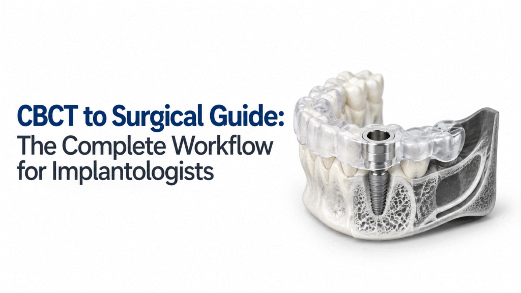
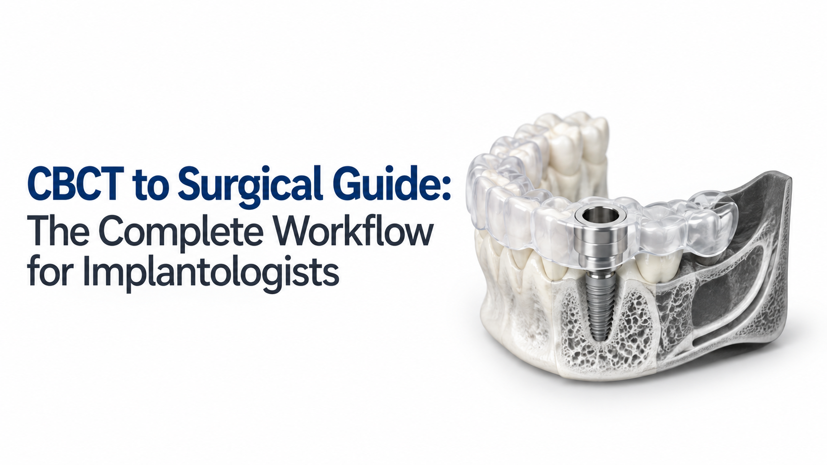
The CBCT to surgical guide workflow is a digital implant-planning process that combines three-dimensional CBCT information with intraoral or surface scan data to plan implant placement and produce a patient-specific surgical guide.
In practical terms, the workflow moves from anatomical records to a digital implant plan and then to a physical guide:
CBCT → intraoral scan → registration → prosthetically driven planning → guide design → clinical review → manufacturing → verification → guided surgery
The important point is that the guide is only one part of the process. Your clinical decisions determine what the guide is designed to transfer, while the quality of the imaging, registration and manufacturing determines how reliably that plan can be transferred.
Step 1: Capture the CBCT and Intraoral Scan
The workflow begins with usable patient data. CBCT provides 3D information about the jaws, available bone, and relevant anatomy. The intraoral scan captures the surface anatomy, teeth, gingiva, and prosthetic environment.
These datasets have different jobs, so both need to be suitable before they are combined.
| Dataset | Primary role | Key checks |
| CBCT / DICOM | 3D anatomical assessment | Coverage, artefacts, image quality and relevant anatomy |
| Intraoral scan / STL | Surface and prosthetic information | Completeness, stitching and occlusion |
| Digital wax-up | Restorative planning | Desired tooth position and emergence |
CBCT should also be justified for the clinical question. Keith Horner, PhD, MSc, BChD, FRCR, FDS, DRD, Odont Dr, Professor at the University of Manchester, described the evidence base as supporting a “restrained approach” to CBCT, limiting its use to situations where conventional imaging does not provide enough information for adequate management.
Step 2: Check the Data Before Registration
Do not assume that having both DICOM and STL files means the case is ready for planning. Review the datasets before merging them.
Check for incomplete intraoral scans, motion or metal artefacts, missing anatomical areas, poor scan stitching and inconsistencies between the digital records and the patient’s clinical situation.
This checkpoint is easy to skip when the workflow is busy, but an error introduced here can travel through every subsequent stage. The supplied research specifically identifies data quality, registration verification and approval responsibility as important workflow checkpoints.
Step 3: Register CBCT With the Intraoral Scan
The CBCT and surface scan are aligned in planning software to create a combined digital representation of the patient.
Registration should then be inspected rather than accepted automatically. Stable reference areas can help with alignment, but you still need to check whether the merged datasets make clinical sense.
This is where CBCT to surgical guide workflow dentistry becomes more than a technical process. Software can complete the registration, but the clinician needs to identify an obvious mismatch before that mismatch influences implant planning.
Step 4: Plan the Implant Around the Restoration and Anatomy
Once the datasets are registered, implant planning can begin.
The planned implant position should account for the intended restoration, available bone, relevant anatomy, implant dimensions, angulation, and the planned surgical approach. A good digital plan answers more than “Can the implant fit here?”
Your review should include:
- Prosthetic emergence and restorative position
- Bone volume and morphology
- Important anatomical structures
- Implant length and diameter
- Angulation and depth
- Drill and surgical access requirements
- The restorative workflow
A 2025 randomized clinical trial involving 30 partially edentulous patients compared guided and freehand implant placement using a 3D-printed guide based on intraoral scan and CBCT data. In that study, the guided group showed lower mean angular, coronal and apical deviations.
That supports the value of guided workflows in the study population, but it should not be interpreted as a guarantee of zero deviation.
Step 5: Design the Surgical Guide
After the implant plan is approved, the surgical guide is designed around the planned implant positions and intended surgical protocol.
The design needs appropriate support and stability, access for the planned instrumentation and compatibility with the implant system and surgical components. Guide design also depends on the clinical situation, including the type of support available.
A guide can be digitally perfect and still be clinically unsuitable if the design does not match the actual surgical workflow. That is why component compatibility and guide support should be reviewed before manufacturing.
Step 6: Review and Approve the Digital Plan
Before the guide is manufactured, the implantologist should review the final plan and guide design.
This is a key responsibility checkpoint. A planning technician, laboratory or external guide provider may prepare the digital workflow, but the treating clinician must decide whether the implant position and proposed guide are clinically appropriate.
| Checkpoint | What should be confirmed? |
| Implant position | Does it meet the restorative objective? |
| Anatomy | Has the relevant anatomy been reviewed in 3D? |
| Registration | Are CBCT and surface data correctly aligned? |
| Guide | Is the support and design appropriate? |
| Components | Are the guide and surgical components compatible? |
| Surgical plan | Does the physical workflow match the digital plan? |
Step 7: Manufacture and Verify the Guide
Once approved, the guide can move to manufacturing, commonly through a 3D-printing workflow.
Quality control should continue after printing. The physical guide should be checked for defects, fit, conformity to the approved design and compatibility with the intended surgical components.
The research dossier includes a 2025 randomized study of 80 implants, with 75 datasets analysed. The study reported measurable differences between two static guided systems, including mean entry-point deviations of 0.64 mm and 0.75 mm and angular deviations of 3.22° and 3.80°.
The practical lesson is simple: guided surgery gives you a controlled way to transfer a plan, but the plan, guide and surgical execution still matter.
Where an External Radiology and Guide Partner Fits
You do not have to perform every stage in-house. A practice may capture the patient’s scan while an external partner handles radiology reporting, digital planning or customized guide production.
The important part is defining ownership at each handoff. Everyone involved should know who captures the data, who interprets the CBCT, who prepares the plan, who designs the guide, who approves it and who checks the final physical guide.
This is one of the clearest gaps identified in the supplied competitor research. Many existing resources explain the technology or production sequence, but fewer explain the dentist-facing handoff between imaging, radiology, planning, and customized guide production.
Nidaan Dental’s verified service ecosystem includes OPG and CBCT imaging, Nidaan Radtech teleradiology and structured reporting by certified Oral & Maxillofacial Radiologists, and Nidaan Uniguide customized surgical guides. It has 40+ years of dental imaging experience and a referral network of 1,200+ dentists.
The Complete CBCT to Surgical Guide Workflow
The complete process can be reduced to:
Acquire → Check → Register → Plan → Design → Review → Manufacture → Verify → Operate
Every stage affects the next. A surgical guide cannot correct poor imaging, incorrect registration or an unsuitable implant plan.
The goal of a digital implant workflow is therefore not automation for its own sake. It is a repeatable process in which the right data reaches the right person, each important decision has a clear owner and the final guide represents the implant plan that the clinician actually approved.
Computer-guided surgery can improve positional control, but published studies still report measurable deviations. The guide should be treated as one part of a controlled clinical workflow, not as a replacement for clinical judgement.
FAQs
1. What files are needed for a CBCT to surgical guide workflow?
Most digital workflows require CBCT DICOM data and an intraoral or surface scan, commonly supplied as STL data. A digital wax-up or restorative plan may also be used when prosthetically driven planning is required. The exact data requirements depend on the planning and guide system being used.
2. Why is CBCT used for surgical guide planning?
CBCT provides three-dimensional information about the jaw and relevant anatomy that can support implant planning. It can be combined with surface-scan information to create a digital representation of the patient’s anatomy and restorative situation. CBCT should still be prescribed based on the clinical need for 3D information.
3. Can a surgical guide eliminate implant placement errors?
No. A surgical guide can help transfer a planned implant position, but it does not eliminate deviation. A 2024 meta-analysis of 67 clinical studies found measurable mean deviations at the entry point, apex, and implant angulation.
4. Who should approve the final surgical guide?
The treating implantologist or responsible clinician should approve the implant plan and determine whether the final guide is appropriate for the intended procedure. A laboratory, software operator, or guide provider can support the technical workflow, but clinical responsibility should remain clearly defined.
5. Can CBCT planning and surgical guide fabrication be outsourced?
Yes. Practices can work with external imaging, radiology, planning, or guide-production partners. The key is to establish clear data requirements, handoff points, review responsibilities and final approval before the guide is manufactured.