

TL;DR
- Inferior alveolar nerve (IAN) injury is one of the most significant complications of mandibular dental implant surgery because it can lead to numbness, tingling, altered sensation, or, in some cases, long-term neuropathic pain.
- CBCT for dental implants allows clinicians to visualize the inferior alveolar nerve, mental foramen, anterior loop, and other anatomical structures in three dimensions before implant placement, helping them make safer treatment decisions.
- However, CBCT is not a guarantee against nerve injury. It is one layer of a comprehensive risk-management workflow that combines accurate diagnosis, careful treatment planning, surgical expertise, and postoperative monitoring.
- A structured planning protocol including clinical assessment, multiplanar nerve tracing, identification of anatomical variations, individualized safety margins, and verification of the surgical guide offers the best protection against avoidable nerve injury.
- Modern implant dentistry is no longer about simply placing an implant where bone is available. It is about placing the right implant in the safest possible position while preserving critical anatomical structures.
Direct Answer
CBCT helps reduce the risk of inferior alveolar nerve injury during dental implant surgery by providing a three-dimensional view of the mandibular canal, mental foramen, anterior loop, and surrounding bone. This detailed visualization enables clinicians to plan implant position, angulation, and depth more accurately than conventional two-dimensional imaging. However, CBCT alone cannot prevent nerve damage. Safe implant placement depends on appropriate case selection, accurate interpretation of the scan, conservative safety margins, careful surgical execution, and continuous clinical judgment throughout treatment.
A practical way to understand it is this:
- CBCT improves visibility.
- Digital planning improves precision.
- Clinical expertise protects the patient.
When these elements work together, implant surgery becomes more predictable and significantly safer.
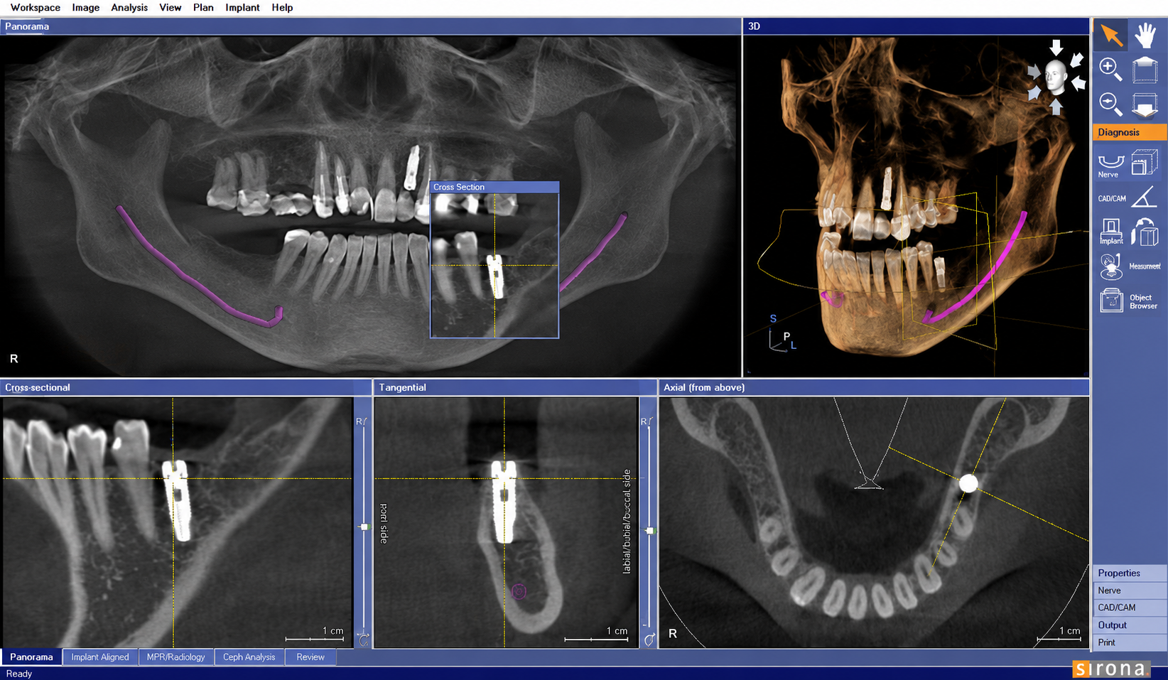
Why Nerve Damage Matters in Dental Implant Surgery
Dental implants have transformed restorative dentistry, but every implant procedure requires careful respect for the patient’s anatomy. In the posterior mandible, one of the most important structures is the inferior alveolar nerve (IAN). Even when an implant integrates successfully with the bone, placing it too close to this nerve can result in sensory complications that may affect a patient’s quality of life.
This is why experienced implantologists spend as much time planning surgery as they do performing it. Modern implant dentistry focuses on preventing avoidable complications before the first drill ever touches the bone.
As Professor Michael M. Bornstein, Professor Keith Horner, and Professor Reinhilde Jacobs, internationally recognized experts in dental radiology, emphasize, CBCT should be viewed as a tool that improves planning rather than a technology that guarantees safety. Their research highlights that evidence does not support the claim that CBCT alone eliminates complications such as nerve injury, reinforcing the need for comprehensive clinical decision-making.
What Is the Inferior Alveolar Nerve (IAN)?
The inferior alveolar nerve is the primary sensory nerve that travels through the mandibular canal. It supplies feeling to several important structures, including:
- The lower teeth
- The lower lip
- The chin
- Part of the gingiva and surrounding soft tissues
Because implants placed in the posterior mandible often lie close to this nerve, even a small planning error can have significant clinical consequences.
What Happens if the Nerve Is Injured?
The severity of nerve injury varies from patient to patient. Some people experience temporary symptoms that improve over time, while others may develop persistent neurosensory problems requiring specialist management.
Possible symptoms include:
- Persistent numbness of the lower lip or chin
- Tingling or “pins and needles” sensations (paresthesia)
- Burning or electric shock-like pain (dysesthesia)
- Altered touch or temperature sensation
- Difficulty speaking or eating comfortably
- In rare situations, long-term neuropathic pain
An important clinical point is that an implant can be technically successful from a surgical perspective while the patient continues to experience significant sensory problems if the nerve has been affected.
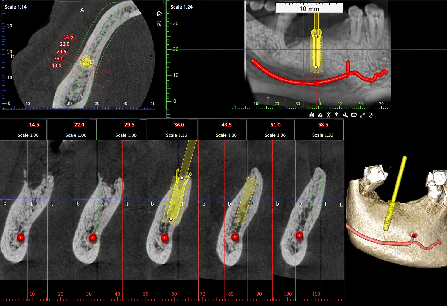
Why CBCT Matters Before Implant Surgery
Planning implant surgery with only a conventional panoramic radiograph can sometimes leave important anatomical details hidden. Two-dimensional images compress complex anatomy into a single flat image, making it difficult to fully appreciate the relationship between the planned implant site and nearby nerves.
CBCT changes this by creating a three-dimensional representation of the patient’s anatomy, allowing clinicians to evaluate implant sites from multiple perspectives before surgery begins.
The Limitations of Traditional 2D X-rays
Conventional panoramic or periapical radiographs remain valuable for routine diagnosis, but they cannot reliably demonstrate several anatomical features that influence implant safety.
These include:
- Buccolingual position of the mandibular canal
- Canal depth within the jawbone
- Anterior loop of the mental nerve
- Accessory mental foramina
- Bifid or trifid mandibular canals
- Lingual concavities
- True cross-sectional bone anatomy
When implants are planned close to these structures, relying solely on two-dimensional imaging may increase diagnostic uncertainty.
How CBCT Improves Implant Planning
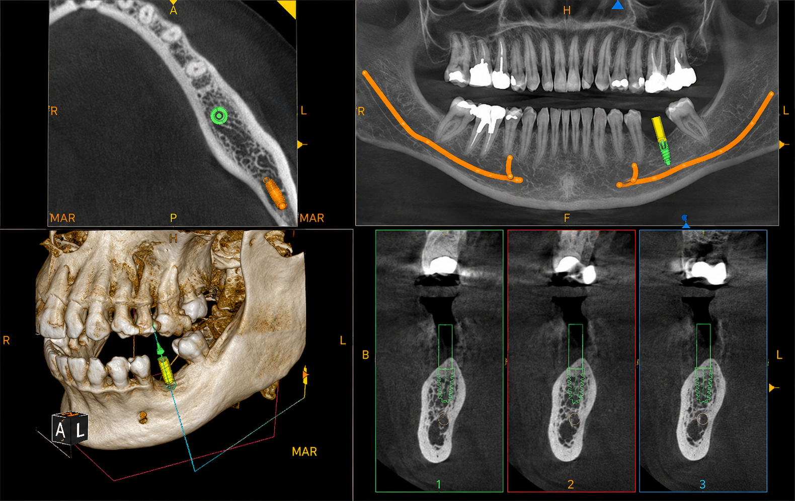
Unlike panoramic imaging, CBCT provides multiplanar visualization through axial, coronal, sagittal, and cross-sectional views. This enables clinicians to understand the patient’s anatomy in three dimensions and evaluate how an implant will relate to surrounding structures before surgery.
CBCT supports clinicians by helping them:
- Measure available bone height and width accurately
- Locate the mandibular canal precisely
- Identify anatomical variations that may alter treatment planning
- Assess implant angulation and trajectory
- Integrate digital planning with surgical guide design
However, professional recommendations remain clear: CBCT should be prescribed only when three-dimensional information is necessary to answer a clinical question, not simply because the technology is available. Appropriate field of view and radiation optimization remain essential parts of responsible imaging.
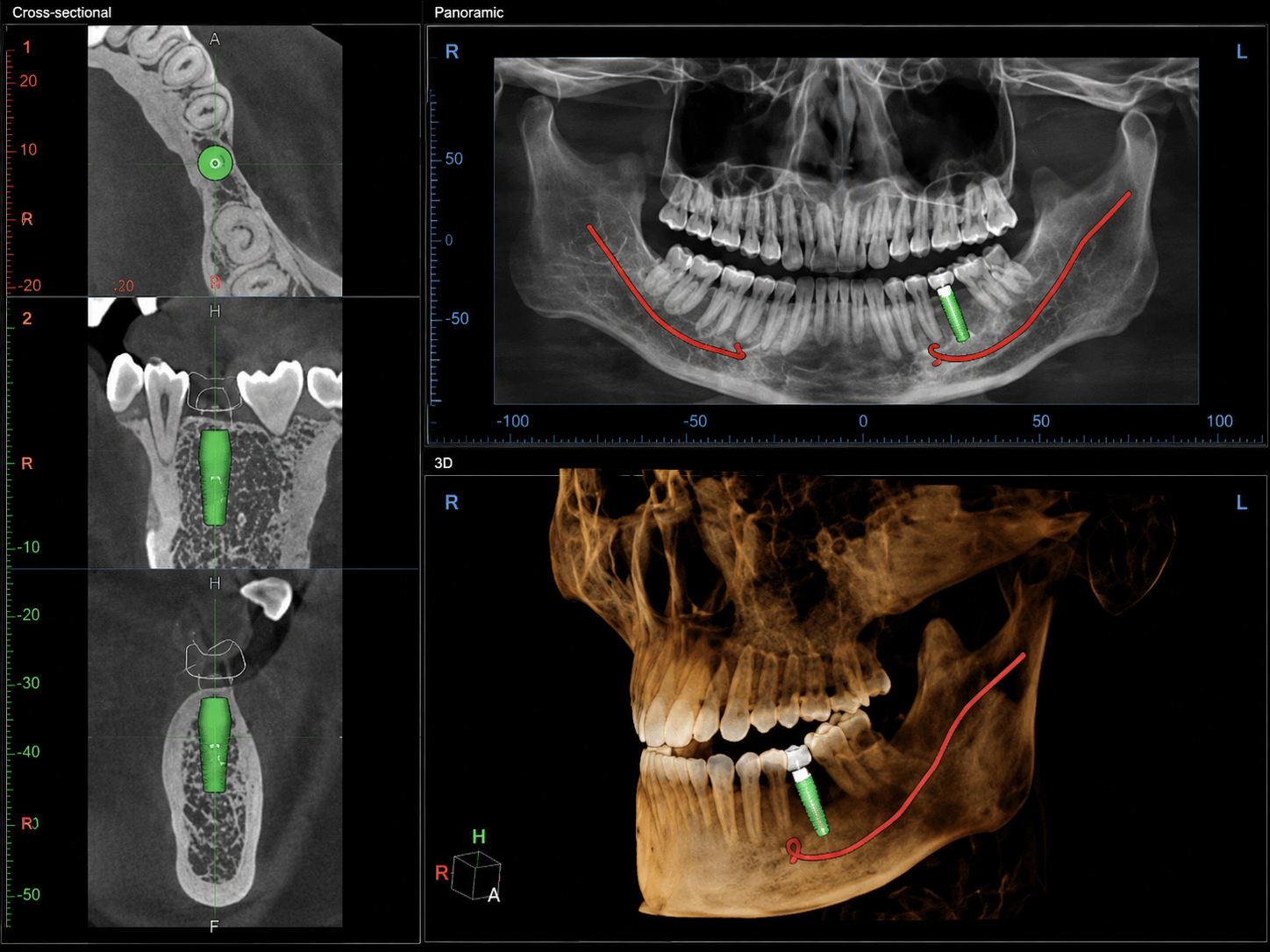
The 7-Step CBCT Safety Workflow Before Implant Surgery
Modern implant planning follows a layered approach to risk reduction rather than relying on a single technology. The uploaded research consistently emphasizes that CBCT delivers its greatest value when incorporated into a structured clinical workflow instead of being treated as a standalone solution.
Step 1: Begin With a Comprehensive Clinical Risk Assessment
Before reviewing the CBCT scan, clinicians first evaluate the patient’s overall clinical situation. Imaging should answer a specific diagnostic question rather than replace clinical examination.
Several factors influence the level of surgical risk, including previous trauma, existing altered sensation, bone availability, implant location, planned implant dimensions, grafting requirements, and immediate loading protocols. When these factors are considered together, the CBCT scan becomes far more valuable because it is interpreted within the appropriate clinical context rather than in isolation.
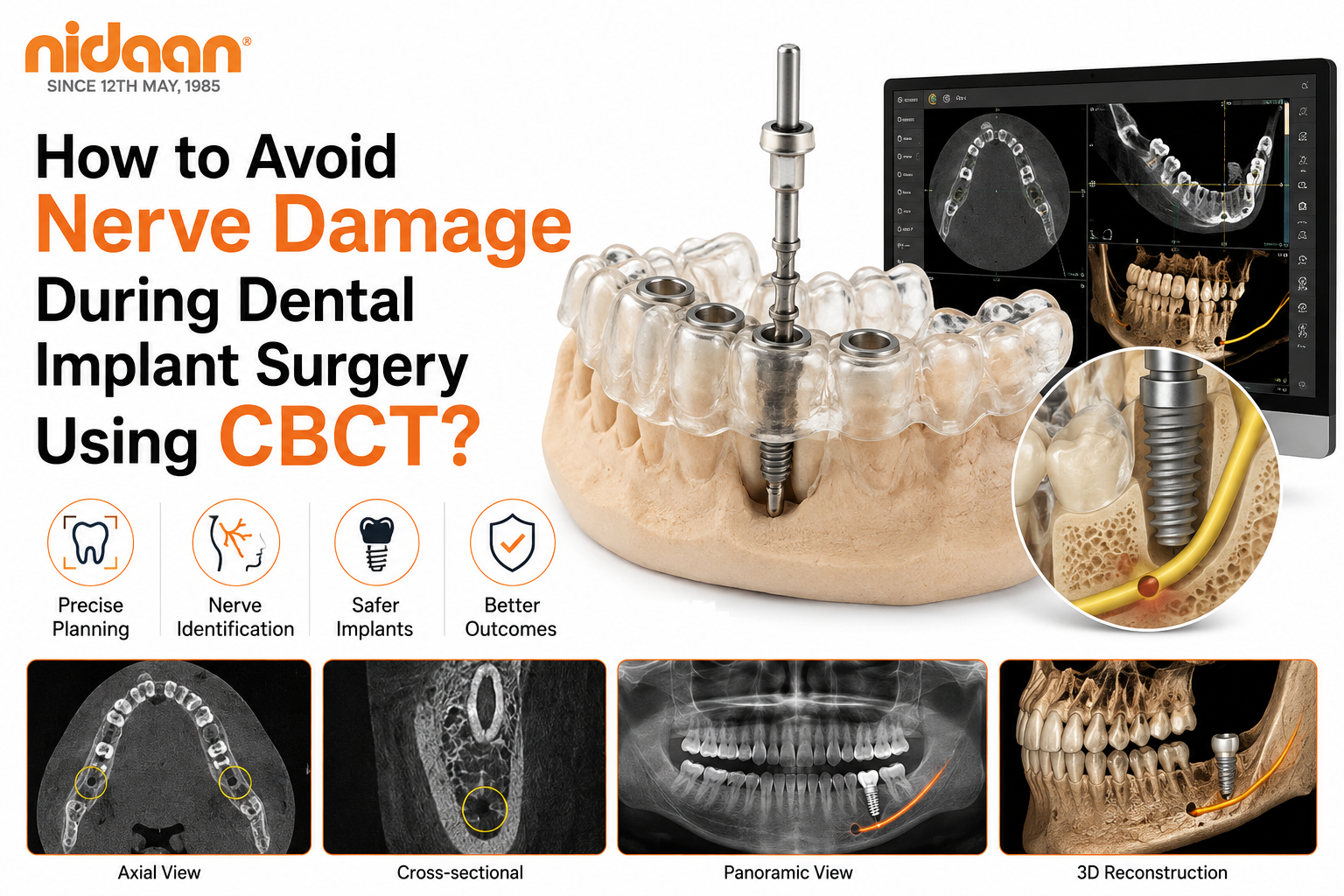
Step 2: Trace the Inferior Alveolar Nerve in Multiple Planes
One of the most important steps in implant planning is accurately identifying the entire course of the inferior alveolar nerve.
Rather than examining a single image, clinicians review the nerve continuously using:
- Axial views
- Coronal views
- Sagittal views
- Cross-sectional slices
- Panoramic reformatted images
Three-dimensional renderings can assist orientation, but they should never replace detailed multiplanar evaluation because subtle anatomical variations may only become visible in cross-sectional analysis. This careful tracing process helps clinicians understand exactly where the nerve lies relative to the planned implant site and supports safer treatment planning.
Step 3: Locate the Mental Foramen Carefully
The mental foramen is the opening through which the mental nerve exits the mandible to supply sensation to the lower lip and chin. Although textbooks often illustrate it in a standard position near the premolars, its actual location varies considerably from one patient to another.
This anatomical variation is one reason why implant planning should never rely on assumptions. Before placing an implant in the premolar region, clinicians carefully evaluate the mental foramen on CBCT to determine its exact location, orientation, and relationship to the proposed implant site.
By identifying the mental foramen in three dimensions, clinicians can modify implant position, length, or angulation when necessary, reducing the likelihood of avoidable nerve injury while preserving prosthetic objectives. This step illustrates an important principle throughout modern implant planning: every patient’s anatomy is unique, and treatment should be individualized accordingly.
Step 4: Evaluate the Anterior Loop
One of the most commonly overlooked anatomical variations is the anterior loop of the mental nerve. Instead of exiting directly through the mental foramen, the nerve may extend forward before looping back toward the foramen. If this extension is not recognized during planning, an implant that appears safe on a panoramic image may actually encroach on the nerve pathway.
CBCT allows clinicians to evaluate the presence, length, and direction of the anterior loop using multiplanar views rather than estimation. Because the extent of this loop varies between individuals, professional recommendations emphasize patient-specific assessment rather than applying a fixed measurement to every case.
Recognizing the anterior loop before surgery often influences implant positioning and helps clinicians maintain a conservative approach in anatomically sensitive regions.
Step 5: Screen for Anatomical Variations
Not every mandibular canal follows the textbook anatomy shown in diagrams. Variations are common, and identifying them before surgery is one of the greatest advantages of three-dimensional imaging.
During CBCT evaluation, clinicians systematically assess for:
- Bifid mandibular canals
- Trifid canals
- Accessory mental foramina
- Retromolar canals
- Lingual foramina
- Lingual concavities
Although some of these structures are uncommon, overlooking them can alter surgical planning. Rather than treating every patient as anatomically identical, CBCT supports a personalized approach where implant positioning reflects the individual’s actual anatomy rather than generalized assumptions.
Step 6: Measure in Three Dimensions
Successful implant planning is not simply about measuring available bone height. Every implant must be evaluated in relation to surrounding anatomical structures across all three spatial dimensions.
Clinicians assess:
- Apical clearance from the mandibular canal
- Buccolingual bone width
- Mesiodistal spacing between adjacent teeth or implants
- Planned implant angulation
- Osteotomy trajectory
- Available restorative space
This comprehensive assessment allows the implant to be planned from the final restoration backward, rather than fitting the restoration around an incorrectly positioned implant. By measuring the implant site three-dimensionally, clinicians reduce uncertainty and improve the predictability of both surgery and the final prosthetic outcome.
Step 7: Verify the Plan Before Surgery
The final planning stage involves verifying every element of the digital workflow before treatment begins.
This includes confirming:
- Inferior alveolar nerve tracing
- Mental foramen location
- Implant diameter and length
- Implant angulation
- Digital planning accuracy
- Surgical guide seating and stability (if guided surgery is planned)
Verification is especially important because every stage of the digital workflow from CBCT acquisition to guide fabrication introduces small tolerances. Reviewing the complete plan before surgery helps ensure that these cumulative variations remain within clinically acceptable limits.
Rather than assuming technology is always correct, experienced clinicians verify every critical detail before proceeding with implant placement.
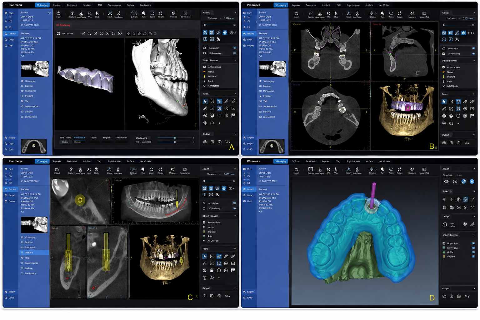
Important Anatomical Structures Evaluated on CBCT
One of the greatest advantages of CBCT implant planning is the ability to evaluate multiple anatomical structures simultaneously. Understanding these landmarks helps clinicians determine where implants can be placed safely while minimizing unnecessary risk.
| Anatomical Structure | Why It Matters During Implant Planning |
| Inferior Alveolar Canal | Identifies the course of the main sensory nerve within the mandible. |
| Mental Foramen | Locates where the mental nerve exits the jaw to avoid injury during premolar implant placement. |
| Mental Nerve | Helps preserve sensation to the lower lip and chin. |
| Incisive Canal | Important when planning implants in the anterior mandible. |
| Anterior Loop | May extend beyond the mental foramen, requiring individualized planning. |
| Accessory Mental Foramina | Anatomical variation that can influence implant positioning. |
| Bifid Mandibular Canal | Additional nerve branches that may not be visible on panoramic imaging. |
| Retromolar Canal | Variant anatomy relevant in posterior mandibular surgery. |
| Lingual Concavity | Helps assess the risk of lingual cortical perforation during osteotomy preparation. |
Because no two patients share identical anatomy, evaluating these structures in three dimensions helps clinicians develop safer, patient-specific treatment plans rather than relying on generalized anatomical expectations.
Understanding Safety Margins Around the Inferior Alveolar Nerve
A common misconception is that maintaining a fixed 2 mm safety margin completely prevents nerve injury. While the literature frequently discusses planning buffers of 1–2 mm, current evidence does not support a single universal distance that guarantees safety for every patient.
Instead, appropriate safety margins depend on multiple variables, including:
- Image quality
- Anatomical variation
- Measurement uncertainty
- Drill tolerances
- Surgical guide accuracy
- Implant design
- Surgical technique
For this reason, experienced implant clinicians use safety margins as clinical planning tools rather than absolute rules. Conservative, case-specific planning remains more reliable than applying the same measurement to every implant site.
As Professor Reinhilde Jacobs, Professor of Oral Radiology and internationally recognized researcher in dentomaxillofacial imaging, has emphasized throughout her work on implant imaging, accurate three-dimensional visualization improves decision-making, but clinical interpretation remains essential because anatomical variation exists in every patient.
How Guided Surgery Supports Nerve Safety
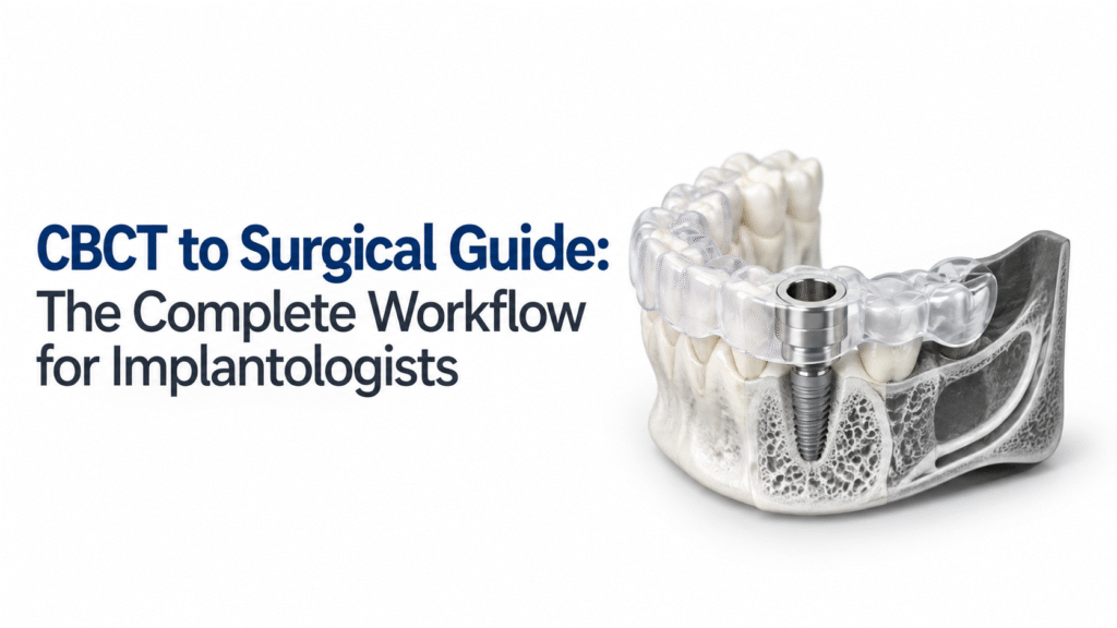
Digital planning does not end with the CBCT scan. In many cases, clinicians combine CBCT data with intraoral scans to create a customized surgical guide that transfers the planned implant position into the patient’s mouth.
When properly designed and verified, guided surgery helps:
- Improve transfer accuracy from digital planning to surgery
- Maintain planned implant angulation
- Control drilling depth and trajectory
- Improve consistency in complex implant cases
However, a surgical guide should never be viewed as the primary safety measure. Its purpose is to support an already well-planned procedure.
Guide seating must always be verified before surgery, and clinicians should be prepared to modify treatment if unexpected clinical findings arise. A guide enhances planning accuracy it does not replace surgical judgment.

Common Planning Mistakes That Increase Nerve Injury Risk
Even with advanced imaging, preventable planning errors can still increase surgical risk. Research consistently highlights several pitfalls clinicians should avoid.
Common mistakes include:
- Relying solely on panoramic imaging for complex mandibular implant cases
- Assuming every patient has “typical” anatomy
- Overlooking the anterior loop of the mental nerve
- Depending entirely on automatic nerve tracing without manual verification
- Measuring only implant length while ignoring angulation and trajectory
- Applying a fixed 2 mm rule regardless of anatomical variation
- Assuming a surgical guide eliminates surgical risk
Avoiding these mistakes requires combining CBCT findings with careful clinical examination, individualized planning, and conservative decision-making throughout the treatment process.
Real Clinical Example
A patient requires replacement of a missing lower second premolar. The panoramic radiograph appears to show adequate bone height for a standard implant. Before surgery, the clinician obtains a CBCT scan.
Three-dimensional evaluation reveals:
- The mental foramen is positioned slightly more anteriorly than expected.
- The anterior loop extends into the originally planned implant site.
- The available clearance to the inferior alveolar canal is less than estimated on the panoramic image.
Instead of proceeding with the original plan, the clinician selects a shorter implant, adjusts the implant angulation, and maintains a more conservative safety margin around the nerve. The implant treatment remains successful not because CBCT prevented nerve injury automatically, but because it revealed critical anatomical information that changed the treatment plan before surgery began.
Myth vs Fact
| Myth | Evidence-Based Fact |
| CBCT completely prevents nerve injury. | CBCT improves three-dimensional visualization and planning but cannot eliminate surgical risk. |
| Guided surgery is always safe. | Surgical guides improve transfer accuracy but cannot compensate for poor planning or inaccurate nerve mapping. |
| A fixed 2 mm safety margin guarantees protection. | Safety margins should be individualized based on anatomy, imaging quality, and surgical factors. |
| Every implant requires CBCT. | CBCT should be prescribed when three-dimensional information is clinically necessary to answer a specific clinical question. |
What the Evidence Actually Supports
Current evidence strongly supports:
- Three-dimensional visualization of the inferior alveolar nerve
- Multiplanar nerve tracing
- Identification of anatomical variations
- Prosthetically driven implant planning
- Selective use of guided surgery
- Conservative, patient-specific safety margins
At the same time, the evidence does not support absolute claims such as:
- “CBCT makes nerve damage impossible.”
- “A universal 2 mm rule is safe for every patient.”
- “Guided surgery guarantees implant success.”
- “Automatic nerve tracing eliminates the need for clinician review.”
These distinctions are important because they reinforce an evidence-based message: technology improves planning, but clinical expertise remains the foundation of safe implant surgery.
Conclusion
CBCT is valuable not simply because it creates a three-dimensional image. Its greatest contribution is helping clinicians make better-informed, patient-specific decisions before implant surgery begins.
When combined with accurate nerve mapping, multiplanar assessment, individualized safety margins, guided surgery verification, careful surgical execution, and postoperative monitoring, CBCT becomes one layer of a comprehensive risk-management strategy.
The most important takeaway is also the simplest: CBCT reduces uncertainty it does not eliminate risk. Safe implant outcomes are achieved when advanced imaging supports sound clinical judgment, not when it replaces it.
Continue Learning About Guided Implant Dentistry
Successful implant treatment begins long before the surgery itself. As you’ve seen throughout this guide, CBCT imaging, digital implant planning, and customized surgical guides work together to improve precision, support prosthetically driven treatment, and help clinicians manage anatomical risks more effectively. However, every patient presents a unique combination of bone anatomy, restorative goals, and clinical considerations, making individualized planning essential.
If you’d like to explore how customized surgical guides are designed and used in modern implant workflows, learn more about Custom Surgical Guides for Implants. The page explains how Nidaan UniGuide integrates CBCT imaging, digital planning, and patient-specific guide fabrication to support accurate implant placement.
You can also visit a Nidaan CBCT & OPG Centre in Pune if your dentist has recommended three-dimensional imaging for implant treatment planning.
Frequently Asked Questions (FAQs)
1. Can a CBCT scan completely prevent nerve damage during dental implant surgery?
No. A CBCT scan significantly improves the clinician’s ability to visualize the inferior alveolar nerve, mental foramen, anterior loop, and surrounding anatomy in three dimensions, making treatment planning much more accurate than conventional two-dimensional imaging. However, CBCT cannot eliminate the risk of nerve injury. Safe outcomes depend on a combination of accurate image interpretation, individualized planning, appropriate safety margins, surgical expertise, and careful execution during the procedure.
2. Why is CBCT preferred over a panoramic X-ray for implant planning near the inferior alveolar nerve?
A panoramic X-ray provides only a two-dimensional overview of the jaws, which can make it difficult to determine the exact position of the mandibular canal or assess bone width accurately. CBCT for dental implants provides multiplanar, three-dimensional views that allow clinicians to evaluate bone dimensions, nerve location, implant angulation, and anatomical variations with much greater precision. This additional information supports safer treatment planning when implants are close to vital structures.
3. What is the inferior alveolar nerve, and why is it important during implant surgery?
The inferior alveolar nerve (IAN) is the main sensory nerve that travels through the lower jaw and provides sensation to the lower teeth, chin, lower lip, and part of the gums. If an implant or surgical instrument comes too close to this nerve, patients may experience numbness, tingling, altered sensation, or, in rare cases, persistent neuropathic pain. Protecting this nerve is therefore one of the most important objectives during mandibular implant planning.
4. Does every dental implant procedure require a CBCT scan?
Not necessarily. Current clinical guidelines recommend prescribing CBCT only when three-dimensional information is necessary to answer a specific clinical question or influence treatment planning. Straightforward implant cases with sufficient anatomical information from conventional imaging may not always require CBCT. Your dentist will recommend the most appropriate imaging based on your individual case and diagnostic needs.
5. What anatomical structures are evaluated on CBCT before placing a dental implant?
During CBCT implant planning, clinicians commonly assess several critical anatomical landmarks, including:
- Inferior alveolar nerve (mandibular canal)
- Mental foramen
- Anterior loop of the mental nerve
- Incisive canal
- Accessory mental foramina
- Bifid or trifid mandibular canals
- Lingual concavities
- Bone height, width, and ridge morphology
Evaluating these structures helps clinicians plan implant placement more accurately while minimizing unnecessary surgical risk.
6. How does guided implant surgery improve safety?
Guided implant surgery uses a patient-specific surgical guide created from CBCT data and digital planning. The guide helps transfer the planned implant position, angle, and drilling path accurately during surgery. While this improves consistency and planning precision, it does not replace careful diagnosis or clinical judgment. A surgical guide should always be considered one part of a comprehensive treatment workflow rather than a guarantee against complications.
7. What is the anterior loop, and why is it important?
The anterior loop is an anatomical extension of the mental nerve that may continue forward before exiting through the mental foramen. Its length and position vary significantly between individuals. If it is not identified during planning, an implant placed in the premolar region could unintentionally affect the nerve. CBCT allows clinicians to evaluate the anterior loop in three dimensions so that implant position can be modified when necessary.
8. Is there a universally safe distance between a dental implant and the inferior alveolar nerve?
No. Although many clinical protocols discuss planning buffers of 1–2 mm, there is no universal safety distance that guarantees nerve protection for every patient. Appropriate safety margins depend on individual anatomy, image quality, implant dimensions, surgical technique, and measurement uncertainty. Experienced clinicians determine safety margins on a case-by-case basis rather than relying on a fixed rule.
9. How does Nidaan UniGuide support safer implant planning?
Nidaan UniGuide combines CBCT imaging, digital implant planning, and customized surgical guide fabrication to help clinicians transfer a carefully planned implant position accurately during surgery. By integrating three-dimensional anatomical information with patient-specific guide design, it supports prosthetically driven implant placement and helps improve planning precision. Like all guided workflows, it complements rather than replaces clinical expertise and sound surgical judgment.
10. What should you ask your implant dentist before undergoing surgery?
Before your implant procedure, consider asking questions such as:
- Do I need a CBCT scan for my implant case?
- How close is my implant site to the inferior alveolar nerve or mental foramen?
- Will my treatment involve a customized surgical guide?
- Is guided implant surgery appropriate for my case?
- How do you evaluate anatomical variations such as the anterior loop?
- What precautions will you take to minimize the risk of nerve injury?
- If unexpected findings occur during surgery, how will the treatment plan be adjusted?
These questions can help you better understand your treatment plan and have an informed discussion with your dentist about the benefits, limitations, and safety considerations of your implant procedure.