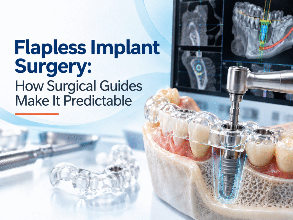

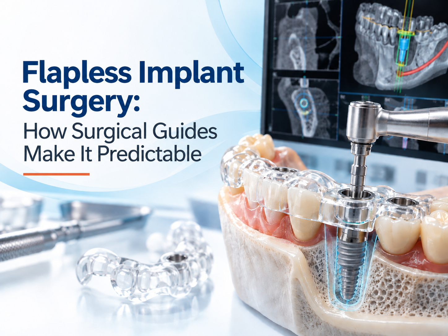
Flapless Implant Surgery: How Surgical Guides Make It Predictable

Dr. Akshay Shah
September 11, 2026
TL;DR
- Flapless implant surgery places the implant without raising a conventional mucoperiosteal flap.
- The technique can reduce surgical exposure, but it also limits direct visual access to the underlying bone.
- A surgical guide transfers a digitally planned implant position to the surgical field.
- CBCT provides three-dimensional information about bone, adjacent roots, the mandibular canal, sinus and other anatomical structures.
- Intraoral scans can be combined with CBCT data to connect the planned implant position with the restorative design.
- Guided surgery can improve placement accuracy and reproducibility compared with freehand placement.
- A 2024 systematic review and meta-analysis of 67 clinical studies reported mean deviations of 1.11 mm at implant entry, 1.40 mm at the apex and 3.51° in angulation.
- Flapless surgery is not suitable simply because a surgical guide is available. Direct access or augmentation may still require flap elevation.
- The guide cannot correct a poor virtual plan. Planning errors, CBCT limitations, scan-registration errors and guide-fit problems can all affect the final result.
- Predictability comes from the complete workflow, not from the printed guide alone.
What Is Flapless Implant Surgery?
Flapless implant surgery involves placing an implant through the soft tissue without raising a conventional surgical flap to expose the underlying alveolar bone.
The attraction is clear. When the clinical situation allows it, avoiding flap elevation can reduce surgical exposure and may contribute to less postoperative swelling and discomfort. It can also support a minimally invasive workflow.
But there is an important trade-off: less surgical exposure means less direct visibility of the bone.
That makes planning particularly important. The clinician needs to know the three-dimensional anatomy before drilling rather than relying on what can be seen during surgery.
This is where CBCT-based guided implantology becomes useful.
Why Is Flapless Implant Surgery More Dependent on Planning?
In freehand surgery, the clinician makes the implant osteotomy while continuously using clinical anatomy, visual access and surgical judgment to guide positioning.
With a flapless approach, direct inspection of the bone is limited. The surgeon therefore relies more heavily on preoperative imaging, the planned implant position and the accuracy with which that plan can be transferred to the patient.
CBCT can show the relationship between the planned implant and structures such as:
- The mandibular canal
- Maxillary sinus
- Adjacent tooth roots
- Cortical plates
- Available ridge dimensions
A digital plan can then establish the intended implant position, angulation and depth before surgery.
The surgical guide acts as the physical connection between that digital plan and the surgical procedure.
How Does a Surgical Guide Make Flapless Surgery More Predictable?
A surgical guide does not make surgery automatically predictable. Its value comes from controlling several parts of the workflow.
1. CBCT Creates the Three-Dimensional Foundation
The process starts with appropriate imaging. CBCT provides three-dimensional information that can be used to assess the proposed implant site and surrounding anatomy.
For example, the plan can account for the available bone and the position of critical structures before surgery begins.
This is particularly relevant in flapless cases because the clinician has less opportunity to directly inspect the bone during the procedure.
2. Intraoral Scans Connect the Implant Plan to the Restoration
CBCT can be combined with an intraoral scan or dental model to create a more complete digital representation of the patient.
This allows implant positioning to be considered alongside the intended tooth position, prosthetic space and restorative requirements.
The objective is not simply to place an implant where bone is available. The implant needs to occupy a position that makes sense biologically and prosthetically.
3. The Implant Position Is Planned Before Surgery
The digital workflow allows the implant’s intended position to be reviewed before the patient enters surgery.
The clinician can assess:
- Implant location
- Angulation
- Depth
- Relationship with neighbouring structures
- Prosthetic position
- Inter-implant relationship in multiple-implant cases
This creates a reference that can be reviewed and adjusted before manufacturing the guide.
4. The Guide Transfers the Planned Position
Once the plan is confirmed, the surgical guide is manufactured to direct the drills according to the planned trajectory.
This is where guided surgery differs from simply using CBCT for diagnosis. The information from the scan becomes part of an actionable surgical workflow.
For multiple implants, this can be particularly useful because the same planned relationship can be transferred across implant sites, helping coordinate implant parallelism, spacing and the prosthetic design.
What Does the Evidence Say About Guided Implant Accuracy?
The evidence supports improved placement accuracy with computer-guided implant surgery, although the results vary between systems, study designs and clinical conditions.
A 2024 systematic review and meta-analysis covering 67 clinical studies reported mean deviations of:
| Measurement | Mean deviation |
|---|---|
| Implant entry point | 1.11 mm |
| Implant apex | 1.40 mm |
| Angular deviation | 3.51° |
These are pooled research findings, not a guarantee of the deviation that will occur in an individual case. Accuracy can be influenced by the support type, guide fixation, sleeves, manufacturing method and the quality of the digital data.
A 2025 randomized trial involving 30 partially edentulous patients also reported lower mean deviations in the guided group than the freehand group. Mean angular deviation was 2.4° ± 1.2° with guidance compared with 6.8° ± 2.1° for freehand placement. Coronal deviation was 0.7 ± 0.3 mm versus 1.9 ± 0.6 mm, while apical deviation was 1.1 ± 0.4 mm versus 2.5 ± 0.9 mm.
The important point is that guidance can reduce variability in implant positioning. It does not mean every guided implant will match the virtual plan exactly.
When Is Flapless Guided Surgery Appropriate?
Flapless surgery should be considered a case-selection decision, not simply a benefit of having a surgical guide.
It may be appropriate when the anatomy has been adequately assessed, the planned implant position is suitable, the soft tissue allows access, and there is no clinical need for direct visualization or simultaneous augmentation.
The research dossier makes an important distinction: a clinician should not use a guide to avoid raising a flap when direct inspection or augmentation is clinically necessary.
This matters because a guide controls the drilling pathway. It does not remove the need to manage the biological condition of the surgical site.
What Can Go Wrong in Guided Flapless Surgery?
A Surgical Guide Cannot Fix a Bad Plan
The most important limitation is simple: the guide transfers the plan. It does not validate the plan by itself.
If the virtual implant is positioned incorrectly, the guide can reproduce that error accurately.
Potential planning problems include inadequate implant-to-tooth spacing, excessive buccal positioning, incorrect implant dimensions, insufficient apical clearance or failure to account for the restorative path.
Digital Registration Can Introduce Errors
The workflow depends on accurate data.
Potential sources of error include:
- Patient movement during CBCT
- CBCT distortion or metal artifacts
- Inaccurate intraoral scans
- Poor scan stitching
- Incorrect merging of CBCT and surface-scan data
- Incorrect identification of anatomical landmarks
That is why quality control matters at every stage rather than only during guide manufacturing.
Flapless Does Not Mean Risk-Free
Guided surgery can help manage the planned implant trajectory, but it cannot eliminate surgical complications.
A safety margin from critical anatomical structures remains necessary. The digital plan should be treated as a highly useful planning reference, not as a perfectly exact representation of what will happen clinically.
From CBCT to Flapless Surgery: The Complete Workflow
A predictable workflow usually follows these stages:
1. Data capture
CBCT and intraoral scan data are collected.
2. Digital planning
The implant position is planned with consideration of anatomy and the restorative objective.
3. Plan review
The dentist reviews the proposed implant position and requests changes where necessary.
4. Guide design
A patient-specific surgical guide is designed from the approved digital plan.
5. Manufacturing and quality control
The guide is fabricated and checked for appropriate fit and compatibility.
6. Surgical execution
The guide is seated correctly and used with the appropriate drilling protocol.
7. Clinical verification
The surgeon remains responsible for confirming the clinical situation and modifying the approach when the intraoperative findings require it.
Nidaan UniGuide follows this type of CBCT-to-guide workflow, combining CBCT and intraoral scan data, digital implant planning, dentist review, customized guide fabrication and delivery with a guided surgery drill kit.
How Nidaan UniGuide Supports Flapless Implant Surgery
Nidaan UniGuide is a customized surgical guide workflow built around CBCT data and intraoral scan information.
The workflow begins with data preparation at the dental clinic. Nidaan’s planning team then works with the digital records to plan the implant position and design the patient-specific guide. The dentist reviews the proposed plan before the guide moves to the printing stage.
The finished UniGuide is delivered with a compatible surgical drill kit for the planned procedure. This gives the dentist a defined digital-to-clinical workflow rather than treating the surgical guide as an isolated product.
For dentists considering flapless implant surgery, this distinction is important. Predictability comes from the quality of the imaging, planning, review, guide design, manufacturing and surgical execution working together.
Conclusion
Flapless implant surgery can be a useful minimally invasive approach when the patient’s anatomy and treatment plan make it appropriate. But removing the flap also reduces direct visibility, which makes accurate preoperative planning more important.
CBCT helps establish the three-dimensional anatomical picture. Intraoral scanning adds restorative information. Digital planning defines the intended implant position, and the surgical guide transfers that plan to the surgical field.
The evidence supports better placement accuracy with guided workflows, but it does not justify treating a surgical guide as a guarantee of clinical success. Data quality, registration, planning, guide fit, case selection and surgical judgment still matter.
For a dentist considering flapless implant surgery, the practical question is not “Can a surgical guide make this case predictable?” It is “Is this case suitable for flapless surgery, and is the entire digital workflow accurate enough to support the planned procedure?”
That is where guided implantology adds real value.
FAQs
1. What is flapless implant surgery?
Flapless implant surgery is an implant placement technique performed without raising a conventional surgical flap. The implant site is accessed through the soft tissue, which can reduce surgical exposure when the case is appropriately selected.
2. How does a surgical guide help with flapless implant surgery?
A surgical guide transfers a digitally planned implant position to the surgical field. It can help control the planned drilling trajectory, implant angulation and depth, reducing variability in implant positioning when the guide is properly designed, fitted and used.
3. Is flapless implant surgery safer than conventional implant surgery?
Not automatically. Flapless surgery can reduce surgical exposure and may support less invasive treatment, but it also provides less direct visibility of the underlying bone. It should only be used when the anatomy and clinical situation support the approach.
4. Does CBCT need to be used for guided flapless implant surgery?
CBCT is commonly used as the three-dimensional imaging foundation for digital implant planning because it provides information about the available bone and surrounding anatomical structures. In a CBCT-based guided workflow, the scan can be combined with intraoral scan data to plan the implant position.
5. How accurate are guided implant surgical guides?
Accuracy varies by system, workflow and clinical conditions. A 2024 systematic review and meta-analysis of 67 clinical studies reported mean deviations of 1.11 mm at implant entry, 1.40 mm at the apex and 3.51° in angulation. These are pooled study results and should not be interpreted as guaranteed accuracy for an individual case.
6. Can a surgical guide eliminate implant placement errors?
No. A guide can reduce variability in transferring a digital plan, but it cannot eliminate errors. Problems can arise from CBCT quality, intraoral scanning, data registration, planning, guide manufacturing, guide seating or surgical execution.
7. Can every implant case be performed flaplessly with a surgical guide?
No. Some cases require direct visualization of the bone, soft-tissue management or augmentation. A surgical guide should not be used simply to avoid flap elevation when clinical access is necessary.
8. What is the difference between guided implant surgery and flapless implant surgery?
They describe different aspects of treatment. Guided implant surgery refers to using a surgical guide to control implant placement according to a digital plan. Flapless surgery refers to the surgical access technique, where a conventional flap is not raised. Guided surgery can be performed with or without a flap, depending on the case.
9. What data are needed to create a custom surgical guide?
A CBCT scan is typically used to provide three-dimensional anatomical information, while an intraoral scan or dental model can provide the surface and restorative information needed for digital planning. Nidaan’s UniGuide workflow uses CBCT together with intraoral scan data for patient-specific guide planning.
10. Does guided flapless surgery guarantee faster healing?
No. A minimally invasive or flapless approach may be associated with less postoperative discomfort or swelling in some situations, but healing depends on several factors, including the surgical procedure, whether grafting is required, the number of implants and individual patient factors. It would be inaccurate to attribute every recovery benefit to the surgical guide alone.