
TL;DR
- Patrick Battiston was knocked unconscious and reportedly lost 2–3 teeth, suffered a fractured jaw, cracked ribs, vertebral injuries, and severe concussion symptoms.
- Tooth loss after high-impact facial trauma is often only one part of a much larger injury pattern.
- Modern sports dentistry would likely use Cone Beam Computed Tomography (CBCT) to evaluate hidden fractures, root injuries, alveolar bone damage, and jaw trauma.
- Studies show CBCT can significantly influence treatment decisions in dental trauma cases, with 84% of endodontists initiating treatment after CBCT review compared with 45% using conventional radiographs alone.
- CBCT has demonstrated approximately 40% higher detection rates for certain traumatic dental injuries compared with conventional imaging methods.
- The Schumacher–Battiston collision highlights a reality modern clinicians understand well: the most serious injuries are often the ones you cannot immediately see.
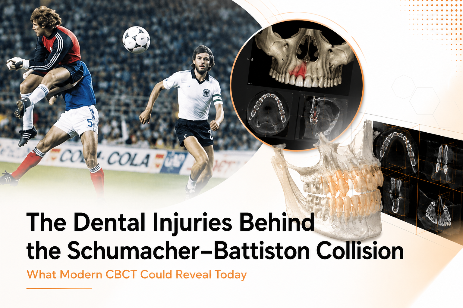
The collision between Harald “Toni” Schumacher and Patrick Battiston during the 1982 FIFA World Cup semi-final remains one of football’s most controversial moments. While the incident is usually discussed as a refereeing controversy, it also represents a significant example of sports-related dental and facial trauma.
Direct Answer
If the Schumacher–Battiston collision happened today, a modern sports dentistry team would likely use CBCT imaging to evaluate the full extent of dental and facial trauma. While conventional dental X-rays can identify obvious tooth injuries, CBCT provides three-dimensional visualization of tooth roots, jawbones, fracture lines, alveolar bone, and surrounding structures. This allows clinicians to diagnose hidden injuries more accurately, develop better treatment plans, and monitor long-term recovery following severe facial trauma.
What If One of Football’s Most Famous Collisions Happened Today?
When football fans remember the 1982 World Cup semi-final between France and West Germany, they usually focus on the controversy.
They remember the collision. They remember Battiston lying motionless on the pitch. They remember the referee allowing play to continue. What rarely gets discussed is dental trauma.
From a modern sports dentistry perspective, the Schumacher–Battiston collision provides a powerful case study in how severe facial impacts affect far more than visible teeth.
When a player loses teeth following a high-speed collision, clinicians are immediately concerned about much more than the missing tooth itself.
The reason is simple. The visible injury is often only the beginning. Modern dental trauma protocols are built around identifying injuries that may remain hidden beneath the gums, inside the jawbone, or within the supporting structures of the teeth.
This is where advanced imaging has fundamentally changed trauma assessment. Today, clinicians have access to diagnostic tools that simply did not exist in routine dental practice in 1982.
One of Football’s Most Controversial Challenges
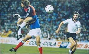
On July 8, 1982, during the FIFA World Cup semi-final in Seville, French defender Patrick Battiston raced onto a through ball before colliding with West German goalkeeper Harald “Toni” Schumacher. The challenge became one of the most infamous incidents in football history.
Historical reports indicate that Battiston:
- Lost 2–3 teeth
- Was knocked unconscious
- Suffered cracked ribs
- Sustained vertebral injuries
- Experienced severe concussion symptoms
- Required oxygen treatment and stretcher evacuation from the field
Remarkably, no foul was awarded. More than forty years later, the incident remains one of the most debated moments in World Cup history. From a clinical standpoint, however, the event offers another lesson. Severe facial trauma rarely affects only the structures that are immediately visible.
Understanding the Dental Trauma Behind the Collision
When someone loses teeth after a major facial impact, dentists immediately begin looking for associated injuries. This is because traumatic dental injuries rarely occur in isolation.
A collision powerful enough to destroy teeth often transfers force into the surrounding bone, supporting tissues, jaw joints, and facial structures.
As the American Academy of Pediatric Dentistry notes, the majority of sport-related dental injuries involve the upper lip, maxilla, and maxillary incisors.
In Battiston’s case, modern clinicians would likely evaluate several categories of trauma simultaneously.
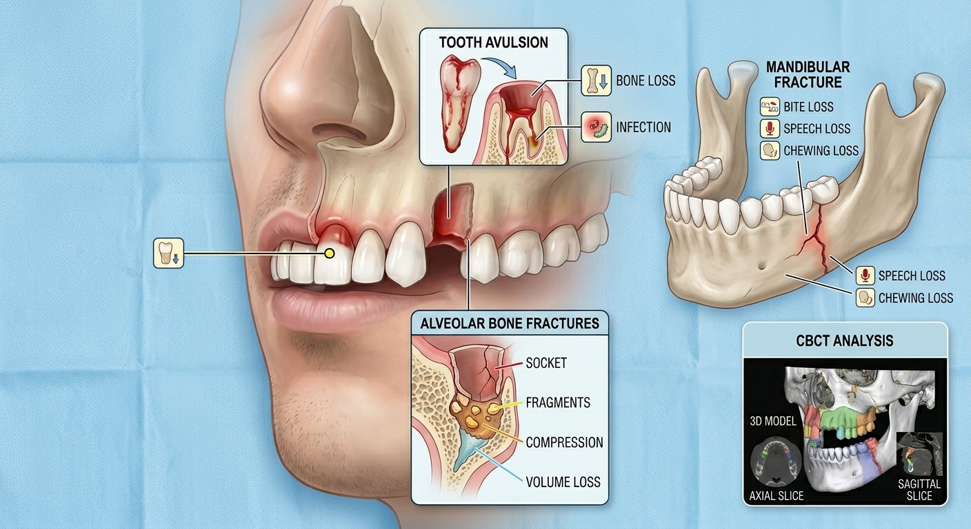
Tooth Avulsion
The most obvious injury would be tooth avulsion, where a tooth is completely displaced from its socket. This represents one of the most urgent emergencies in sports dentistry because treatment timing significantly affects long-term outcomes.
Current estimates suggest that more than 5 million teeth are avulsed annually in the United States alone, creating an annual treatment burden exceeding $500 million.
For athletes, immediate diagnosis and management are critical because complications can include:
- Root resorption
- Bone loss
- Infection
- Future implant requirements
Alveolar Bone Fractures
A tooth does not simply sit inside the mouth. It is anchored within the alveolar bone. When a collision is strong enough to knock teeth out, there is a substantial possibility that the surrounding bone has also been damaged.
This may include:
- Socket wall fractures
- Bone fragmentation
- Bone compression
- Loss of supporting bone volume
These injuries can significantly affect future restorative treatment and are often difficult to evaluate accurately using conventional radiographs alone.
Mandibular and Facial Fractures
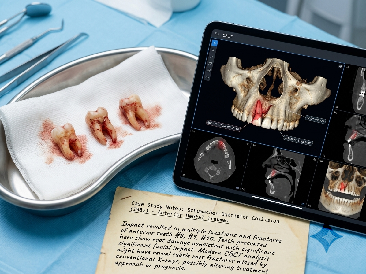
Historical reports indicate Battiston suffered a fractured jaw.
Mandibular fractures can affect:
- Bite alignment
- Speech
- Chewing function
- Facial symmetry
- Long-term oral health
Determining the exact location and severity of these fractures is one of the situations where CBCT can provide substantial diagnostic value.
What Would Today’s Trauma Protocol Look Like?
If Battiston presented to a modern sports dentistry team today, the evaluation would extend far beyond visual inspection. Current trauma protocols focus on identifying both visible injuries and hidden structural damage.
Initial assessment would likely involve:
Clinical Examination
The first stage would evaluate:
- Airway stability
- Neurological status
- Facial symmetry
- Tooth mobility
- Occlusal changes
- Soft tissue injuries
However, clinical examination alone cannot reveal everything. That is where advanced imaging becomes essential.
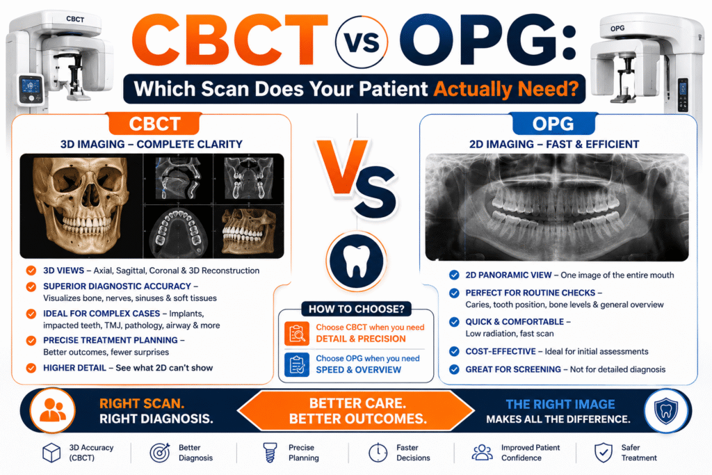
The Role of CBCT Imaging
Modern trauma teams increasingly rely on CBCT because it provides a three-dimensional view of the structures affected by injury.
Unlike conventional dental radiographs, CBCT allows clinicians to examine:
- Teeth
- Root structures
- Alveolar bone
- Jaw fractures
- Facial anatomy
- Joint structures
As researchers from National Library of Medicine have noted:
“Reviewing cone-beam computed tomography (CBCT) scans may influence how endodontists approach treatment decisions in dental trauma cases.”
This matters because treatment decisions often change once hidden injuries become visible.
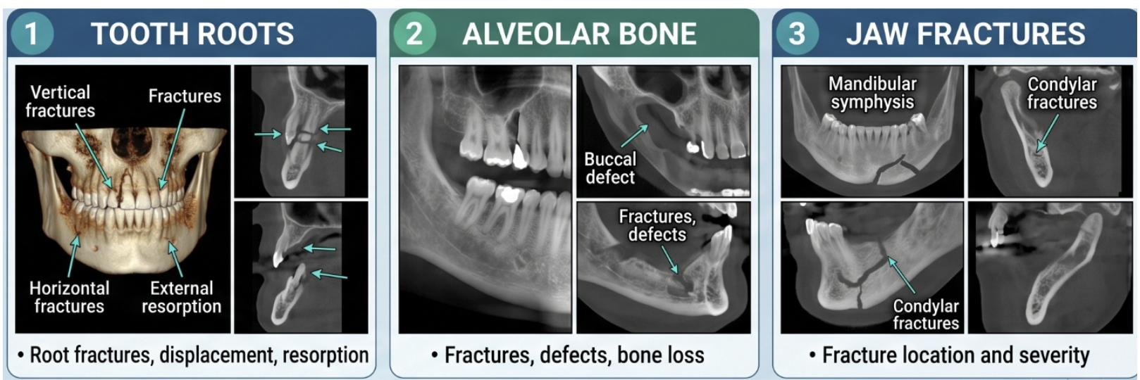
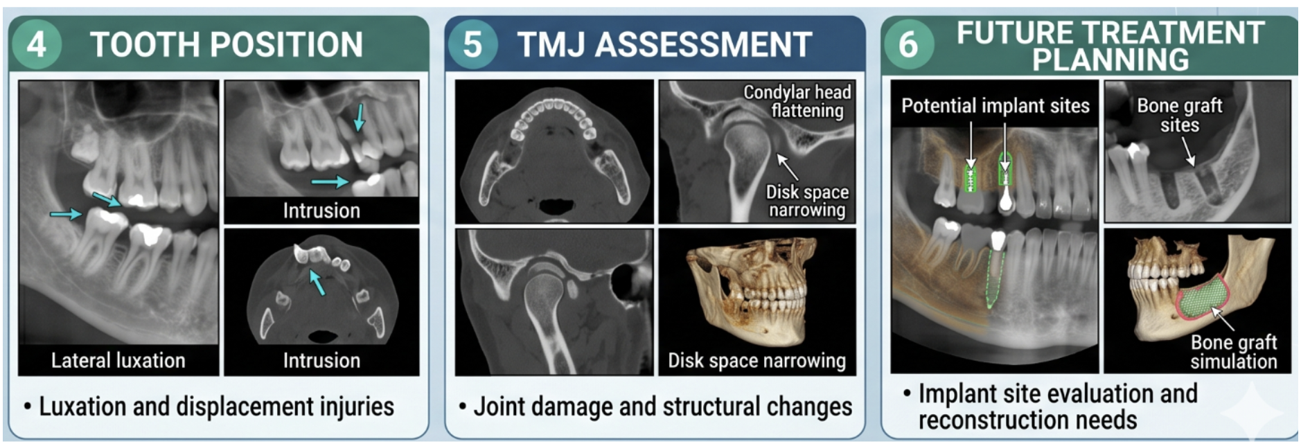
What Would Clinicians Look for on a CBCT Scan?
One of the biggest advantages of CBCT is its ability to reveal injuries that may not be visible during routine examination.
Following a collision like Schumacher–Battiston, clinicians would likely evaluate:
| Trauma Assessment Area | What CBCT Can Reveal |
| Tooth Roots | Root fractures, displacement, resorption |
| Alveolar Bone | Fractures, defects, bone loss |
| Jaw Fractures | Fracture location and severity |
| Tooth Position | Luxation and displacement injuries |
| TMJ Assessment | Joint damage and structural changes |
| Future Treatment Planning | Implant site evaluation and reconstruction needs |
Research published in recent years suggests CBCT demonstrates approximately 40% higher detection rates for certain traumatic dental conditions compared with conventional imaging methods.
That difference can significantly influence treatment planning. In fact, studies cited in current dental literature found that 84% of endodontists initiated treatment after reviewing CBCT scans compared with only 45% when relying on conventional radiographs alone.
The implication is clear. Better visualization often leads to different clinical decisions.
Why This Incident Still Matters in Modern Sports Dentistry
The Schumacher–Battiston collision is often remembered as a football controversy. For dental professionals, it also represents something else. It illustrates how severe sports trauma can affect multiple structures simultaneously and why comprehensive diagnosis matters.
Modern research continues to show that sports-related dental injuries remain common.
Recent studies report:
- Sports account for 11.3–42.1% of facial fractures.
- Approximately 31% of maxillofacial injuries involve dental trauma.
- Contact sports-related oro-dental trauma prevalence can reach 35.9%, while mouthguard use remains surprisingly low at only 20.8%.
These statistics reinforce an important reality. The type of injury Battiston experienced in 1982 is not simply a historical event.
Similar injuries continue to occur in sports today. The difference is that clinicians now have better tools to diagnose and manage them.
From Football History to Modern Trauma Diagnosis
The Schumacher–Battiston collision remains one of football’s most controversial moments, but it also serves as a compelling example of severe sports-related dental trauma.
Battiston reportedly lost multiple teeth and suffered significant facial injuries following the impact. Today, those injuries would likely trigger a far more comprehensive diagnostic process involving advanced imaging technologies such as CBCT.
Modern CBCT allows clinicians to evaluate root fractures, alveolar bone injuries, jaw fractures, tooth displacement, and long-term complications with far greater accuracy than was possible in 1982.
The broader lesson extends beyond football history. A missing tooth may be the injury everyone notices. But in major facial trauma, the injuries that matter most are often the ones hidden beneath the surface.
Learn More About Dental Trauma, CBCT & Advanced Dental Imaging
Severe dental injuries often involve far more than a chipped tooth or visible tooth loss. As the Schumacher–Battiston collision demonstrates, high-impact facial trauma can affect tooth roots, jawbones, supporting structures, and long-term oral health in ways that are not always immediately obvious.
If you’re interested in learning more about:
- CBCT scans in dentistry
- Dental trauma diagnosis
- Sports-related facial injuries
- Implant planning after tooth loss
- OPG vs CBCT imaging
- Modern dental radiology
explore the latest educational resources and diagnostic services available through Nidaan Dental.
Visit the Nidaan Blog for more evidence-based articles on dental imaging, CBCT technology, and oral health. Explore Best CBCT and OPG Scan in Pune to learn more about advanced dental imaging, radiologist-reviewed reporting, and modern diagnostic workflows.
Accurate diagnosis is often the first step toward better treatment outcomes.
Frequently Asked Questions (FAQs)
What dental injuries did Patrick Battiston reportedly suffer after the Schumacher collision?
Historical reports indicate that Patrick Battiston suffered multiple serious injuries following the 1982 World Cup collision, including the loss of approximately 2–3 teeth, a fractured jaw, cracked ribs, vertebral injuries, and severe concussion symptoms. He was reportedly knocked unconscious and required oxygen treatment before being removed from the field on a stretcher.
From a modern dental trauma perspective, these visible injuries would likely prompt clinicians to investigate for additional hidden damage involving the teeth, jawbones, and surrounding facial structures.
What is tooth avulsion and why is it considered a dental emergency?
Tooth avulsion occurs when a tooth is completely knocked out of its socket due to trauma. It is one of the most serious dental emergencies because the long-term survival of the tooth often depends on how quickly treatment is provided.
When a tooth is avulsed, several complications can occur:
- Damage to the periodontal ligament
- Loss of supporting bone
- Root resorption
- Infection
- Permanent tooth loss
Modern dental trauma protocols emphasize immediate evaluation because early intervention can significantly improve the chances of saving the affected tooth.
How would a modern dentist assess an injury like Battiston’s today?
Today, a severe sports-related facial injury would typically be evaluated through a combination of clinical examination and advanced imaging.
The assessment may include:
- Neurological evaluation
- Examination of facial symmetry
- Bite analysis
- Tooth mobility assessment
- Soft tissue examination
- CBCT imaging when clinically indicated
The goal is not only to identify visible injuries but also to detect hidden fractures, bone damage, root injuries, and joint complications that may not be obvious during an initial examination.
Why would CBCT be useful after a sports-related facial injury?
CBCT (Cone Beam Computed Tomography) provides three-dimensional imaging of the teeth, jawbones, roots, and surrounding structures.
After significant facial trauma, CBCT can help clinicians evaluate:
- Root fractures
- Alveolar bone fractures
- Jaw fractures
- Tooth displacement
- Impacted fragments
- Bone loss
- Temporomandibular joint (TMJ) injuries
Unlike conventional dental X-rays, CBCT allows clinicians to examine injuries from multiple angles, improving diagnostic accuracy and treatment planning.
Can CBCT detect injuries that traditional dental X-rays may miss?
Yes. One of the major advantages of CBCT is its ability to identify hidden injuries that may not be visible on conventional two-dimensional radiographs.
Research cited in current dental trauma literature indicates that CBCT can provide significantly improved detection of certain traumatic dental conditions and may influence treatment decisions in a substantial percentage of trauma cases.
This is particularly important when evaluating:
- Root fractures
- Alveolar bone damage
- Complex jaw fractures
- Tooth luxation injuries
- Internal structural defects
For severe trauma cases, this additional diagnostic information can have a direct impact on treatment outcomes.
What types of dental injuries commonly occur during sports?
Sports-related dental trauma can range from minor injuries to complex facial fractures.
Common injuries include:
- Tooth avulsion (knocked-out teeth)
- Crown fractures
- Root fractures
- Tooth displacement (luxation)
- Soft tissue injuries
- Jaw fractures
- TMJ injuries
- Alveolar bone fractures
Research suggests that sports account for a significant proportion of facial injuries worldwide, making sports dentistry an increasingly important field within modern dental care.
Why are root fractures difficult to diagnose without advanced imaging?
Root fractures occur below the visible portion of the tooth and often cannot be detected through visual examination alone.
In many cases:
- The tooth may appear intact
- Symptoms may be delayed
- Conventional X-rays may not capture the fracture line clearly
CBCT allows clinicians to evaluate the tooth from multiple perspectives, increasing the likelihood of identifying fractures that could otherwise remain undiagnosed. Early diagnosis is important because untreated root fractures can lead to infection, pulp necrosis, and eventual tooth loss.
How does CBCT help when planning treatment after tooth loss?
When a tooth cannot be saved following trauma, long-term treatment may involve dental implants or other restorative options.
CBCT plays an important role by allowing clinicians to assess:
- Bone height
- Bone width
- Bone density
- Socket integrity
- Anatomical limitations
This information helps determine whether implant placement is possible and whether additional procedures such as bone grafting may be required.
Accurate imaging improves treatment predictability and helps clinicians create a more personalized treatment plan.
What is the connection between sports dentistry and CBCT technology?
Sports dentistry focuses on the prevention, diagnosis, and treatment of oral and facial injuries in athletes.
Because many sports injuries involve multiple structures simultaneously, modern sports dentists increasingly rely on advanced imaging technologies such as CBCT to obtain a complete understanding of trauma.
CBCT can assist with:
- Diagnosing complex injuries
- Evaluating fractures
- Monitoring healing
- Planning reconstruction
- Supporting long-term follow-up care
This makes it a valuable tool in the management of moderate to severe sports-related dental trauma.
Could a mouthguard have reduced the severity of injuries like Battiston’s?
While no protective device can eliminate all risk, research consistently shows that properly fitted mouthguards significantly reduce the likelihood and severity of dental injuries during contact sports.
Studies cited in current sports dentistry literature suggest:
- Mouthguards may reduce sports-related dental trauma by as much as 82–93%.
- Athletes who do not wear mouthguards may face a 1.6–1.9 times higher risk of orofacial injury.
Organizations involved in sports dentistry continue to advocate for greater awareness and adoption of custom-fitted mouthguards, particularly in high-impact sports.
What is the biggest lesson modern dentistry can learn from the Schumacher–Battiston incident?
The biggest lesson is that visible injuries rarely tell the entire story.
A missing tooth may attract immediate attention, but severe facial trauma can also involve:
- Hidden root fractures
- Jaw fractures
- Bone damage
- Joint injuries
- Long-term complications
Modern dentistry increasingly focuses on understanding the full extent of trauma rather than simply treating the most obvious injury.
Advanced imaging technologies such as CBCT have become valuable because they allow clinicians to see beyond what is visible and make more informed treatment decisions. In many trauma cases, the most important injuries are not the ones you notice first, they are the ones hidden beneath the surface.