


Region: Maxillary Left Posterior (Site 26)
Implants: Two implants, maxillary left posterior
Procedure: Crestal Sinus Lift + Simultaneous Implant Placement
Guide: Nidaan UniGuide (Static, Tooth-Supported)
CASE OVERVIEW: Clinical Presentation & Diagnosis
A partially edentulous patient presented to Nidaan Dental with missing dentition in the maxillary left posterior region, requiring implant-supported rehabilitation. Two dental implants were planned to replace the missing teeth, restoring function and aesthetics in a prosthetically driven approach.
Preoperative evaluation included high-resolution cone beam computed tomography (CBCT) of the maxillary posterior region. Volumetric analysis revealed a critical anatomical constraint at the 26 implant site: inadequate residual bone height secondary to maxillary sinus pneumatization, the process by which the maxillary sinus expands inferiorly, reducing the vertical bone available between the alveolar crest and the sinus floor.
The available bone height at site 26 was insufficient to accommodate a standard-length implant without encroaching on or perforating the Schneiderian membrane, the delicate mucosal lining of the maxillary sinus. To achieve the vertical bone height necessary for safe, stable implant placement, a crestal approach sinus lift (transalveolar sinus floor elevation) was planned simultaneously with implant placement, a technically demanding procedure requiring precise osteotomy depth control and atraumatic sinus floor elevation.
“When anatomy limits us, digital planning and guided surgery expand what is safely possible without compromising on the prosthetic outcome.”
Clinical Summary
| Parameter | Clinical Detail | Status |
|---|---|---|
| Patient | Partially edentulous, maxillary left posterior | ✓ Assessed |
| Missing Teeth | 26 region (maxillary left second premolar / first molar) | ✓ Treated |
| CBCT Finding | Inadequate residual bone height and sinus pneumatization at 26 | ✓ Planned |
| Sinus Procedure | Crestal approach sinus lift (simultaneous with implant) | ✓ Executed |
| Implant Count | Two implants in the maxillary left posterior region | ✓ Placed |
| Surgical Guide | Nidaan UniGuide static tooth-supported guide | ✓ Utilised |
CLINICAL CHALLENGES
Why This Case Demanded Guided Precision
The combination of sinus pneumatization, inadequate bone height, and the need for simultaneous sinus floor elevation and implant placement created a convergence of anatomical and procedural challenges that made freehand surgery inappropriate. Each variable compounded the risk of the next.
| Challenge | Clinical Significance |
|---|---|
| Sinus Pneumatization at 26 | CBCT revealed inadequate residual bone height at the 26 site due to maxillary sinus expansion, placing the planned implant apex at risk of sinus membrane encroachment or perforation without vertical augmentation. |
| Crestal Sinus Lift Risk | The crestal (transalveolar) approach to sinus floor elevation relies on controlled, incremental osteotome pressurisation. Without guided depth control, the risk of Schneiderian membrane perforation, the most critical complication, is substantially elevated. |
| 3D Implant Positioning | Simultaneous sinus lift and implant placement require precise mesiodistal, buccopalatal, and axial positioning. Any deviation from the prosthetically driven plan compromises emergence profile, crown-to-implant ratio, and long-term loading vectors. |
| Anatomical Limitation | The combined constraint of limited bone height and proximity to the sinus floor left minimal margin for error. Even a 1–2mm deviation in depth or angulation could negate the sinus augmentation effect or risk implant exposure into the sinus cavity. |
“In the posterior maxilla, the margin between a predictable outcome and a sinus complication can be less than a millimetre. Guided surgery transforms that margin from a surgical gamble into a planned, controlled parameter.”
The crestal sinus lift approach, while less invasive than the lateral window technique, is critically dependent on controlled, incremental osteotome advancement. A surgical guide that fixes both the entry point and the maximum safe depth translates clinical risk into a measurable, manageable parameter. Without guidance, depth is estimated; with the Nidaan UniGuide, depth is prescribed.
NIDAAN’S SOLUTION
Precision-Guided Protocol: UniGuide + Crestal Sinus Lift
Nidaan Dental’s approach to this case centred on the principle that surgical precision begins at the planning stage, not at the point of incision. A fully integrated digital workflow was employed, from CBCT acquisition and prosthetic planning through to the fabrication and clinical verification of the Nidaan UniGuide, ensuring every anatomical constraint was resolved before the patient entered the surgical chair.
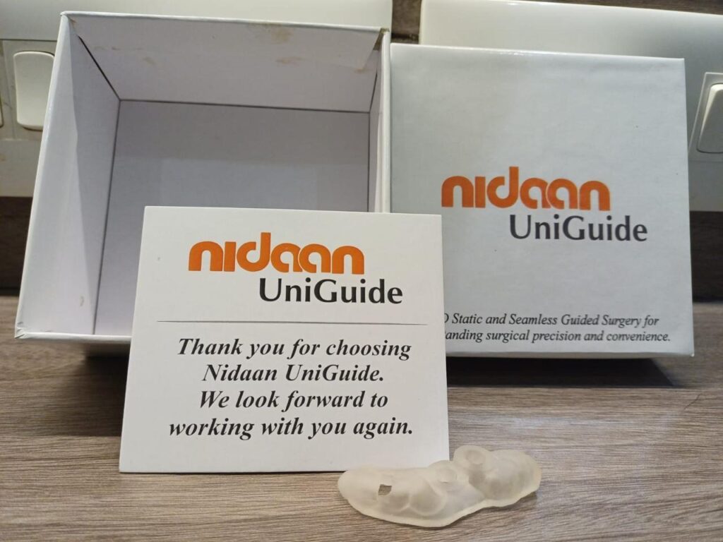
Nidaan UniGuide 3D static tooth-supported surgical guide with branded packaging
| NIDAAN UNIGUIDE SURGICAL GUIDE USED IN THIS CASE: Nidaan UniGuide is a 3D static, tooth-supported surgical guide system providing seamless guided implant surgery with submillimetre accuracy in angulation, depth, and positioning. Why UniGuide was critical for this sinus case: • Controlled osteotomy depth prevents Schneiderian membrane perforation during crestal sinus lift |
Surgical Protocol Step by Step
Step 1: CBCT Analysis & Prosthetic-Driven Implant Planning
High-resolution CBCT data was imported into implant planning software alongside intraoral scan data. The sinus floor was precisely mapped at the 26 site, and the available bone height quantified. The implant position was established from the prosthetic outcome; backward crown position, emergence profile, and abutment axis were defined first, then the implant body was virtually placed within the anatomical envelope. The crestal sinus lift osteotomy depth and graft volume were calculated based on the delta between available bone and required implant length.
Step 2: Nidaan UniGuide Fabrication & Pre-Surgical Verification
The Nidaan UniGuide was designed with tooth-supported retention, eliminating reliance on soft tissue or bone contact for stability, particularly important in the posterior maxilla where tissue compressibility can introduce guide displacement. Drilling sleeves were positioned and angulated to correspond precisely with the planned implant axes. The guide incorporated depth stop calibration for both the osteotomy preparation and the crestal sinus elevation instrument sequence. Fabricated from biocompatible, autoclavable resin via 3D printing, the guide was verified for passive, stable seating before surgery.
Step 3: Flap Elevation & Guided Crestal Osteotomy
Following local anaesthesia and mucoperiosteal flap elevation, the Nidaan UniGuide was seated and confirmed stable. Guided sequential drilling was initiated through the UniGuide sleeves each drill diameter controlled in entry point, angulation, and maximum depth. The depth stop system prevented the osteotome from advancing beyond the planned sinus floor elevation level, providing an inbuilt safeguard against membrane perforation at every stage of the protocol.
Step 4: Crestal Sinus Floor Elevation
With the osteotomy prepared under full guided control, graduated osteotomes were used to incrementally elevate the sinus floor via the crestal approach, the Summers technique, refined with guided entry precision. The Schneiderian membrane was elevated atraumatically with controlled pressure, creating the supra-sinus space required for graft material placement. The guided depth control at the osteotomy stage provided confidence that the membrane had not been perforated before elevation commenced. Particulate bone graft (xenograft or synthetic as indicated) was condensed into the elevated sinus space to support the implant apex and facilitate long-term bone formation.
Step 5: Simultaneous Implant Placement
Following sinus floor elevation and graft condensation, both implants were placed in the maxillary left posterior region through the Nidaan UniGuide. Insertion torque was confirmed at the planned subcrestal depth, with the implant apex engaging the augmented sinus space apically and the available native cortical bone laterally. The guide ensured that throughout placement, from initial seating to final torque, the implant trajectory remained identical to the digital plan. Interimplant parallelism was verified and confirmed.
Step 6: Wound Closure & Post-Operative Protocol
Following implant placement and guide removal, the surgical site was irrigated thoroughly. Flap repositioning and primary closure were achieved. Healing abutments or cover screws were placed as indicated by primary stability values. Post-operative CBCT was available for plan-to-outcome comparison. A structured recall protocol was initiated: week 1 (suture removal and tissue assessment), month 1 (soft tissue healing evaluation), month 3–4 (radiographic osseointegration check), and month 6 (prosthetic phase commencement).
“The Nidaan UniGuide eliminated the two greatest risks of this procedure simultaneously: sinus membrane perforation and implant malposition, by converting both into planned, measurable parameters controlled before the first instrument was placed.”
CONCLUSION
Guided Surgery as the Solution to Posterior Maxillary Complexity
This case demonstrates the clinical value of the Nidaan UniGuide in one of implant dentistry’s most anatomically unforgiving scenarios: simultaneous crestal sinus lift and implant placement in the posterior maxilla with limited residual bone height.
The key determinants of success in this case Schneiderian membrane integrity, precise osteotomy depth, correct implant angulation, and optimal platform positioning relative to the augmented sinus floor were each directly controlled by the UniGuide system. What would have required real-time intraoperative estimation under freehand conditions was instead resolved digitally, before surgery commenced.
The prosthetically driven planning workflow ensured that the implants were positioned not simply where anatomy permitted, but where restoration demanded, achieving an outcome that supports long-term functional loading, crown proportions, and maintenance access.
At Nidaan Dental, guided surgery is not a specialty service reserved for complex presentations. It is our clinical standard applied wherever precision, patient safety, and predictable outcomes matter. In the posterior maxilla, with its proximity to the sinus, its reduced bone volume, and its prosthetic demands, that standard is non-negotiable.