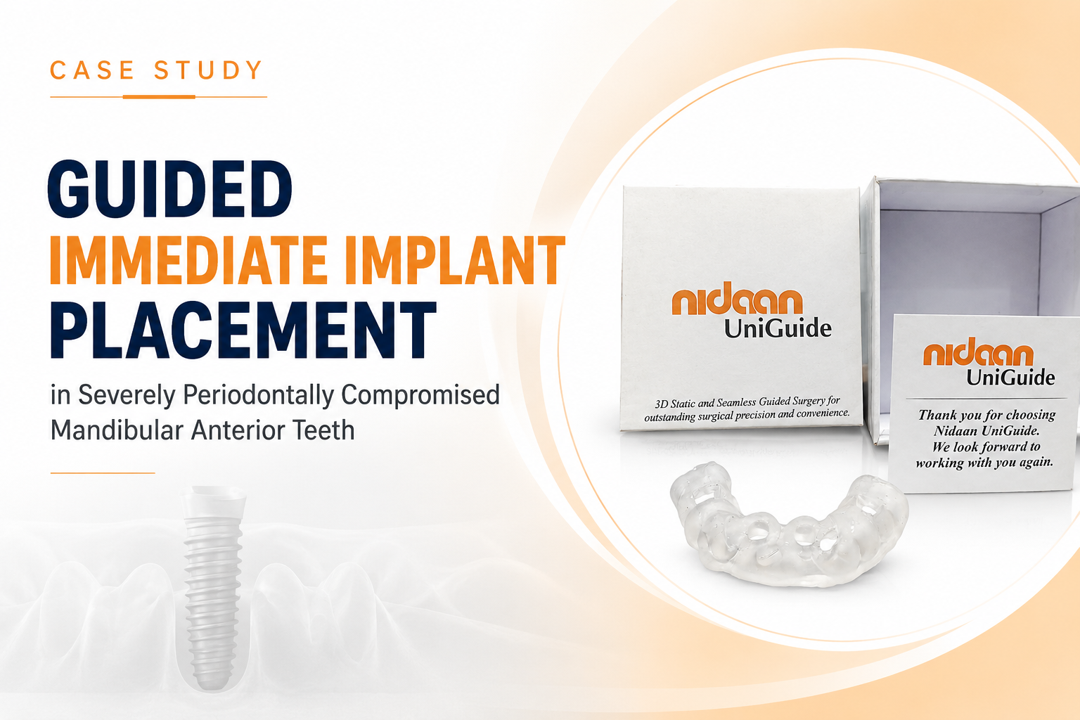
Guided Immediate Implant Placement in Severely Periodontally Compromised Mandibular Anterior Teeth

Dr. Akshay Shah
May 26, 2026
Implant Sites: 32 & 42 (Mandibular Central Incisors)
Protocol: Immediate Placement (Type 1) · Guided Surgery
Surgical Guide: Nidaan UniGuide (Tooth-Supported)
Speciality: Implant Dentistry & Digital Planning
CASE OVERVIEW
Clinical Presentation & Diagnosis
This case presents a patient with advanced periodontal disease affecting the mandibular anterior sextant, resulting in severe bone loss, mobility, and a markedly compromised prognosis for the existing natural teeth at positions 3 2 (lower left central incisor) and 4 2 (lower right central incisor). Clinical and radiographic evaluation confirmed deep probing depths, furcation involvement, and vertical bone defects inconsistent with tooth retention under any viable regenerative protocol.
Following multidisciplinary assessment, a treatment plan was formulated for the extraction of the compromised teeth with simultaneous, immediate implant placement, a protocol demanding exceptional precision given the proximity of adjacent roots, the critical aesthetic importance of the mandibular anterior zone, and the significant anatomical challenges posed by the existing bone architecture.
“Immediate implant placement in the aesthetic zone, particularly in a periodontally compromised environment, demands a level of three-dimensional accuracy that freehand surgery simply cannot consistently deliver.”
The mandibular anterior region presents unique prosthetic and biological demands. The labial plate is typically thin, the interradicular space between adjacent implants is narrow, and the aesthetic expectations, smile line, emergence profile, and crown proportions are unforgiving. Any deviation in implant angulation, depth, or mesiodistal positioning at the time of surgery can cascade into irreversible prosthetic compromise. This clinical reality made computer-guided surgery not merely advantageous but clinically essential.
Clinical Summary
| Parameter | Clinical Finding | Status |
| Periodontal Bone Loss | Severe, bilateral at 32 & 42 | Addressed |
| Tooth Prognosis | Hopeless, indicated for extraction | Resolved |
| Implant Protocol | Immediate placement (Type 1) | Executed |
| Aesthetic Zone Risk | High labial concavity & thin tissue biotype | Mitigated |
| Surgical Guidance | Nidaan UniGuide (tooth-supported) | Utilised |
CLINICAL CHALLENGES
Operating within the mandibular anterior region following periodontal bone loss introduces a convergence of anatomical, biological, and prosthetic variables that collectively raise the surgical risk profile considerably. This case presented four distinct, interconnected challenges, each of which directly influenced the treatment design and reinforced the necessity of a fully guided surgical protocol.
| Challenge | Clinical Significance |
| Severe Labial Concavity | Chronic resorption secondary to periodontal disease created a pronounced undercut morphology. Freehand drilling risked cortical perforation and uncontrolled angulation, directly threatening aesthetic outcomes. |
| Compromised Residual Bone | Advanced horizontal and vertical bone loss reduced available volume for primary implant stability. Achieving adequate insertion torque (>25 Ncm) required engagement of apical and palatal cortical regions. |
| Parallel Placement at Adjacent Sites | Simultaneous implants at 32 and 42 required precise parallelism for prosthetic passivity and adherence to the critical >3mm inter-implant spacing rule to prevent inter-implant bone loss. |
| High-Aesthetic Zone Demands | The mandibular anterior directly influences the social smile. Any deviation in implant depth or labiopalatal inclination would necessitate compromised emergence profiles and potential visible tissue changes. |
“Immediate implant placement carries an inherently higher risk of malposition compared to delayed protocols. In the aesthetic zone, this risk is compounded; the post-extraction socket anatomy rarely aligns with the ideal prosthetic axis, making surgical guidance the single most impactful risk-reduction strategy available.”
Beyond the anatomical complexity, the psychological and clinical consequences of failure in this zone must be acknowledged. Visible implant failure, mid-facial recession, or prosthetic misalignment in the anterior mandible carries significant patient impact. This case demanded a protocol where execution error was eliminated, not merely minimised.
NIDAAN’S SOLUTION
At Nidaan Dental, the treatment philosophy centres on eliminating surgical guesswork through digital planning and fabrication precision. For this case, a comprehensive digital workflow was employed, from cone beam CT acquisition through to the fabrication and clinical verification of the Nidaan UniGuide surgical guide, ensuring that every critical variable was resolved at the planning stage, not intraoperatively.
NIDAAN UNIGUIDE: THE SURGICAL GUIDE USED IN THIS CASE
Nidaan UniGuide is a 3D static, tooth-supported surgical guide system engineered for seamless guided implant surgery, delivering outstanding precision, angulation control, and depth management in a single sterile, CAD/CAM-fabricated appliance.
Key features utilised in this case:
- Tooth-supported design for maximum intraoperative stability without bone or mucosal contact
- Precision sleeve positioning controls entry point, angulation, and depth simultaneously
- Fully guided protocol compatible with sequential drilling systems
- Biocompatible, sterilisable resin fabricated via 3D printing from CBCT + digital scan data
If you want, learn more about Nidaan Uniguide.
Surgical Protocol: Step by Step
Step 1: CBCT-Based 3D Treatment Planning
High-resolution cone beam computed tomography (CBCT) was acquired and co-registered with intraoral scan data. The DICOM dataset was imported into implant planning software for precise volumetric bone mapping. Available bone height, width, and density at 32 and 42 were quantified. The ideal implant position, dictated by the planned prosthetic emergence profile, was established first (prosthetic-driven planning), with the implant body then virtually positioned within the available bone envelope accordingly.
Step 2: Nidaan UniGuide Design & Fabrication
A Nidaan UniGuide tooth-supported surgical guide was selected as the optimal guide typology for this case, providing maximum stability without requiring mucosal or bone contact, which can be unreliable in the presence of significant resorption. The guide was meticulously designed using CAD software to accommodate the planned extraction sockets, incorporate appropriately angulated sleeves for both 3 2 and 4 2, and allow unobstructed access for irrigation and visual confirmation throughout surgery. The guide was fabricated from biocompatible, sterilisable resin via 3D printing.
Step 3: Pre-Surgical Guide Verification
Before surgery, the guide fit was verified on the study model and confirmed clinically for passive, stable seating. Guide stability was assessed under lateral loading to confirm the absence of rocking, a critical pre-operative check ensuring intraoperative accuracy is maintained. Depth stops were calibrated to the planned implant platform position relative to the anticipated post-extraction crestal level.
Step 4: Atraumatic Extraction & Socket Preparation
Periotome-assisted atraumatic extraction was performed at 3 2 and 4 2, prioritising preservation of the residual labial plate. The post-extraction sockets were thoroughly debrided and irrigated. Socket architecture was assessed, confirming Type 1/2 classification (Hammerle & Chen), with the labial concavity anatomically consistent with the preoperative CBCT findings. The UniGuide was seated with stability confirmed before commencement of osteotomy preparation.
Step 5: Fully Guided Osteotomy & Implant Placement
Sequential guided drilling was performed through the Nidaan UniGuide sleeves using the implant system’s guided-surgery drill kit. The fully guided protocol controlled entry point, angulation, and depth simultaneously, negating the labial deflection risk introduced by the cortical concavity. Implants at 3 2 and 4 2 were placed with confirmed primary stability, achieving planned apical anchorage in the subapical cortical bone. Interimplant parallelism was verified clinically and confirmed to match the digital plan. The gap between the implant surface and the labial socket wall was grafted with particulate xenograft to support volume maintenance during early osseointegration.
Step 6: Prosthetic Space Optimisation & Follow-up Protocol
Implant platform depth was confirmed at 3–4mm subcrestal, within the biologically and prosthetically optimal range for anterior implants requiring adequate emergence profile development. Healing abutments or provisional restorations were placed as indicated to support soft tissue contour and mucosal seal. A structured recall protocol was established for osseointegration monitoring, tissue evaluation, and staged restoration delivery.
“The Nidaan UniGuide did not change the biology; it ensured that every decision made during the planning phase was executed with submillimetre fidelity at the surgical site. That is its irreplaceable value.”
CONCLUSION
This case demonstrates that computer-aided surgical guidance, delivered through the Nidaan UniGuide system, transforms high-risk, anatomically demanding immediate implant scenarios into reproducible, predictable outcomes. The convergence of severe periodontal compromise, labial concavity, multi-unit simultaneous placement, and aesthetic zone demands would have made freehand surgery at best unpredictable, at worst contraindicated.
The Nidaan UniGuide resolved each of these variables in a single, integrated planning step, ensuring that the most critical decisions (implant position, angulation, depth, parallelism) were made with three-dimensional data, not intraoperative estimation. The result is not only a surgically successful implant placement, but one in which optimal prosthetic space, emergence profile, and long-term aesthetic stability were built into the plan from day one.
The clinical outcomes achieved in this case reflect the core Nidaan Dental treatment philosophy: precision planning, executed flawlessly. For patients with compromised bone, thin tissue biotypes, or high aesthetic demands, computer-guided implant surgery is not a premium add-on; it is the appropriate standard of care.
As the evidence base for guided protocols continues to mature, the clinical community’s understanding is increasingly clear: in the aesthetic zone, accuracy is not optional. It is the treatment itself. At Nidaan Dental, we remain committed to integrating the highest standards of digital dentistry with individualised clinical care, ensuring that every patient receives not only a functional implant, but a beautiful, biologically stable, long-lasting result.