2D IMAGING
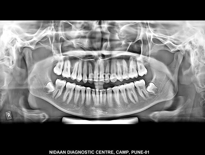
OPG (Orthopantomogram)
An OPG is a panoramic or wide view x-ray of the lower face, which displays all the teeth of the upper and lower jaw on a single film. It demonstrates the number, position and growth of all the teeth including those that have not yet surfaced or erupted. It is different from the small close up x-rays dentists take of individual teeth. An OPG may also reveal problems with the jawbone and the joint which connects the jawbone to the head, called the Temporomandibular joint or TMJ. An OPG may be requested for the planning of orthodontic treatment, for assessment of wisdom teeth or for a general overview of the teeth and the bone which supports the teeth.
Procedure

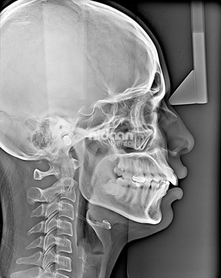
Lateral Cephalogram
A Lateral Cephalogram (Lat Ceph) is a lateral or side view x-ray of the face, which demonstrates the bones and facial contours in profile on a single film. Lat Ceph x-rays are usually used in the diagnosis and treatment of orthodontic problems.
Procedure
You may be asked to remove jewellery, eyeglasses, and any metal objects that may obscure the images. You will be asked to stand with your head against the machine so that it can be adjusted for your comfort. Pair of cone shaped plastic supports are then gently positioned in each ear, rather like a pair of headphones. This aligns both ears to ensure that an exact side view of the face is obtained. You will not feel any discomfort during the procedure.

Frontal Cephalogram (P-A Cephalogram)
A frontal cephalogram is a radiograph of the head taken with the x-ray beam perpendicular to the patient’s coronal plane with the x-ray source behind the head and the film cassette in front of the patient’s face. P-A cephalograms are usually taken for evaluation and treatment planning of patients with facial asymmetry.
Procedure
You may be asked to remove jewellery, eyeglasses, and any metal objects that may obscure the images. You will be asked to stand with your face against the cassette or sensor so that it can be adjusted for your comfort. Pair of cone shaped plastic supports are then gently positioned in each ear, rather like a pair of headphones. This aligns both ears to ensure that an exact view of the face is obtained. You will not feel any discomfort during the procedure.

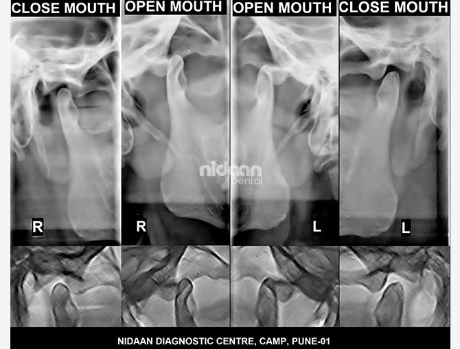
Both Temporomandibular Joint (Both TMJ)
Temporo-mandibular joint (TMJ) disorders are medical problems related to the jaw joint, which connects the jaw or mandible to the temporal bone at each side of the head. As you open and close your mouth each time you talk, chew, sing or yawn, your jaw condyles glide along the joint socket of the temporal bone. Standard x-rays of the TMJs are taken in the open and closed position for each side. Additional, advanced medical imaging, such as Cone Beam CT (CBCT) might also be needed to fully assess the TMJs.
Procedure
You may be asked to remove jewellery, eyeglasses, and any metal objects that may obscure the images. You will be asked to stand with your face resting on a small shelf and to bite on a sterile mouth piece to steady your head. It is important to stay very still while the x-ray is taken. After the first rotation of the x-ray tube around your head, you will be asked to open your mouth as wide as possible for the second rotation. You will not feel any discomfort during the procedure.

